. 2008 Jun 13;283(24):16895-905.
doi: 10.1074/jbc.M800747200. Epub 2008 Mar 14.

Phosphorylation at Ser-129 but not the phosphomimics S129E/D inhibits the fibrillation of alpha-synuclein

Affiliations

Phosphorylation at Ser-129 but not the phosphomimics S129E/D inhibits the fibrillation of alpha-synuclein

Katerina E Paleologou et al. J Biol Chem. .

Abstract

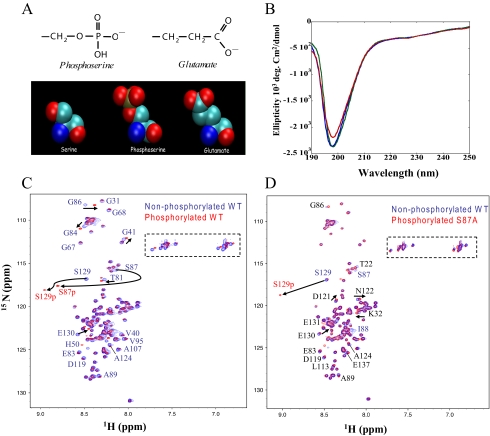
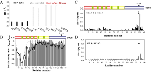
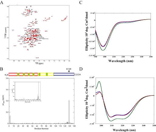
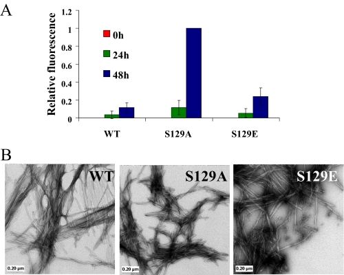
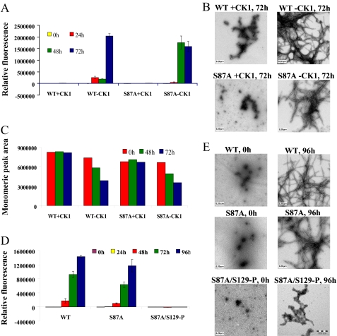
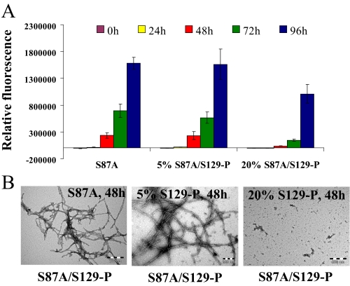
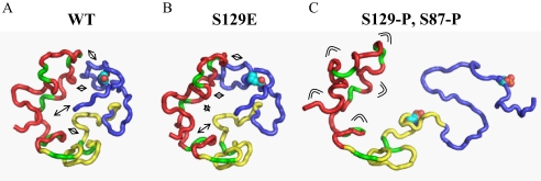
alpha-Synuclein (alpha-syn) phosphorylation at serine 129 is characteristic of Parkinson disease (PD) and related alpha-synulceinopathies. However, whether phosphorylation promotes or inhibits alpha-syn aggregation and neurotoxicity in vivo remains unknown. This understanding is critical for elucidating the role of alpha-syn in the pathogenesis of PD and for development of therapeutic strategies for PD. To better understand the structural and molecular consequences of Ser-129 phosphorylation, we compared the biochemical, structural, and membrane binding properties of wild type alpha-syn to those of the phosphorylation mimics (S129E, S129D) as well as of in vitro phosphorylated alpha-syn using a battery of biophysical techniques. Our results demonstrate that phosphorylation at Ser-129 increases the conformational flexibility of alpha-syn and inhibits its fibrillogenesis in vitro but does not perturb its membrane-bound conformation. In addition, we show that the phosphorylation mimics (S129E/D) do not reproduce the effect of phosphorylation on the structural and aggregation properties of alpha-syn in vitro. Our findings have significant implications for current strategies to elucidate the role of phosphorylation in modulating protein structure and function in health and disease and provide novel insight into the underlying mechanisms that govern alpha-syn aggregation and toxicity in PD and related alpha-synulceinopathies.


Figures

FIGURE 1.
FIGURE 1.
α-Syn is disordered independent of phosphorylation. A, the atomic structures of the side chains of Glu and phospho-Ser demonstrate the structural and electrostatic similarities between the two moieties. B, CD spectra of WT (blue line), S129A (green line), and S129E (red line) α-syn (10 μm). C, comparison of two-dimensional 1H,15N HSQC spectra of unphosphorylated WT (blue) and phosphorylated WT (red) α-syn. D, comparison of two-dimensional 1H,15N HSQC spectra of unphosphorylated S87A (blue) and phosphorylated S87A (red) α-syn. Resonance assignments are indicated with residue numbers. A dashed rectangle marks Gln and Asn side-chain resonances.
FIGURE 2.
FIGURE 2.
Phosphorylation disrupts long-range interactions in monomeric α-syn. A, hydrodynamic radii of various α-syn mutants in phosphate buffer at 15 °C with and without 8 m urea. B, comparison of paramagnetic broadening of amide protons between unphosphorylated WT α-syn (black) and phosphorylated S87A α-syn (gray). In both cases the paramagnetic MTSL spin label was attached to residue 18, which was mutated from Ala to Cys. HSQC spectra in the presence (paramagnetic) and absence (diamagnetic) of spin label were recorded at 15 °C, and the intensity ratio of the resonance peaks was determined. Dashed lines indicate paramagnetic effects expected for a random coil polypeptide. C and D, normalized weighted average 1H,15N chemical shift differences between unphosphorylated and phosphorylated S87A α-syn (C) and between WT and S129D α-syn (D).
FIGURE 3.
FIGURE 3.
Phosphorylation at Ser-129 does not alter the structure of micelle-bound α-syn. A, comparison of two dimensional 1H,15N HSQC spectra of unphosphorylated (black) and phosphorylated (red) S87A α-syn bound to detergent micelles. B, normalized weighted average chemical shift differences between phosphorylated and unphosphorylated S87A α-syn in its micelle-bound state. C, CD spectra of WT (blue line), S87A (green line), and S87A/Ser(P)-129 (red line) α-syn (10 μm). D, circular dichroism spectroscopy of WT (blue line), S87A (green line), and S87A/Ser(P)-129 (red line) α-syn in the presence of POPG vesicles. α-syn, POPG mass ratio is 1:20.
FIGURE 4.
FIGURE 4.
S129E exhibits similar in vitro aggregation properties as the WT protein, but S129A forms significantly more fibrils than WT. A, ThT fluorescence was monitored at indicated time points from samples of 100 μm solutions of WT, S129A, and S129E α-syn incubated at 37 °C. The error bars represent the S.D. of four independent experiments. B, TEM images of WT, S129A, and S129E α-syn after 48 h of aggregation (scale bar, 0.20 μm).
FIGURE 5.
FIGURE 5.
CK1-mediated phosphorylation at Ser-129 inhibits the fibrillation of WT and S87Aα-syn. A, ThT fluorescence measurements of CK1-phosphorylated WT and S87A α-syn and their unphosphorylated controls (100 μm); results are the average of three readings ± S.D. B, TEM images of phosphorylated and unphosphorylated S87A α-syn after 72 h of aggregation at 37 °C (scale bar, 0.20 μm). C, graph representing the area of monomeric peak after 0, 48, and 72 h of aggregation at 37 °C. D, ThT fluorescence measurement of WT, S87A, and S87A/Ser(P)-129 α-syn as a function of time (average of three readings ± S.D.). E, TEM images of WT, S87A, and S87A/Ser(P)-129 α-syn at 0 h and after 96 h of aggregation at 37 °C (scale bar, 0.20 μm).
FIGURE 6.
FIGURE 6.
S87A/Ser(P)-129 interacts with S87A and inhibits its aggregation in a dose-dependent manner. S87A α-syn (70 μm) was mixed with the appropriate volume of 70 μm S87A/Ser(P)-129 to generate samples containing 5 and 20% of the latter protein. A, ThT fluorescence measurement of the 70 μm S87A, S87A containing 5% S87A/Ser(P)-129 α-syn and S87A containing 20% S87A/Ser(P)-129 α-syn (average of 3 readings ± S.D.). B, TEM images of S87A and S87A containing 5 or 20% S87A/Ser(P)-129 after 48 h of aggregation at 37 °C (scale bar, 0.20 μm).
FIGURE 7.
FIGURE 7.
Schematic illustration of the disruption of long-range interactions in α-syn upon phosphorylation at Ser-129. A, representative member of the native state ensemble of conformations populated by α-syn (17). The basic N-terminal region, the highly amyloidogenic NAC region, and the acidic C-terminal domain are shown in red, yellow, and blue, respectively. The side chain of Ser-129 is shown. Long-range interactions are indicated by arrows. B, phosphomimics do not mimic the structural consequences of phosphorylation. Same as B, but the side chain at position 129 was replaced by a Glu. C, upon phosphorylation at Ser-129, long-range interactions are disrupted, and the ensemble of conformations is extended. The higher flexibility of the N-terminal domain is indicated.

References

    1. Trojanowski, J. Q., and Lee, V. M. (2003) Ann. N. Y. Acad. Sci. 991107 -110 - PubMed
    1. Abeliovich, A., Schmitz, Y., Farinas, I., Choi-Lundberg, D., Ho, W. H., Castillo, P. E., Shinsky, N., Verdugo, J. M., Armanini, M., Ryan, A., Hynes, M., Phillips, H., Sulzer, D., and Rosenthal, A. (2000) Neuron 25239 -252 - PubMed
    1. Cooper, A. A., Gitler, A. D., Cashikar, A., Haynes, C. M., Hill, K. J., Bhullar, B., Liu, K., Xu, K., Strathearn, K. E., Liu, F., Cao, S., Caldwell, K. A., Caldwell, G. A., Marsischky, G., Kolodner, R. D., Labaer, J., Rochet, J. C., Bonini, N. M., and Lindquist, S. (2006) Science 313324 -328 - PMC - PubMed
    1. Outeiro, T. F., and Lindquist, S. (2003) Science 3021772 -1775 - PMC - PubMed
    1. Kahle, P. J., Neumann, M., Ozmen, L., and Haass, C. (2000) Ann. N. Y. Acad. Sci. 920 33-41 - PubMed

Publication types