. 2008 May;20(5):659-69.
doi: 10.1093/intimm/dxn025. Epub 2008 Mar 14.

Gr1+IL-4-producing innate cells are induced in response to Th2 stimuli and suppress Th1-dependent antibody responses

Affiliations

Gr1+IL-4-producing innate cells are induced in response to Th2 stimuli and suppress Th1-dependent antibody responses

Amy S McKee et al. Int Immunol. 2008 May.

Abstract

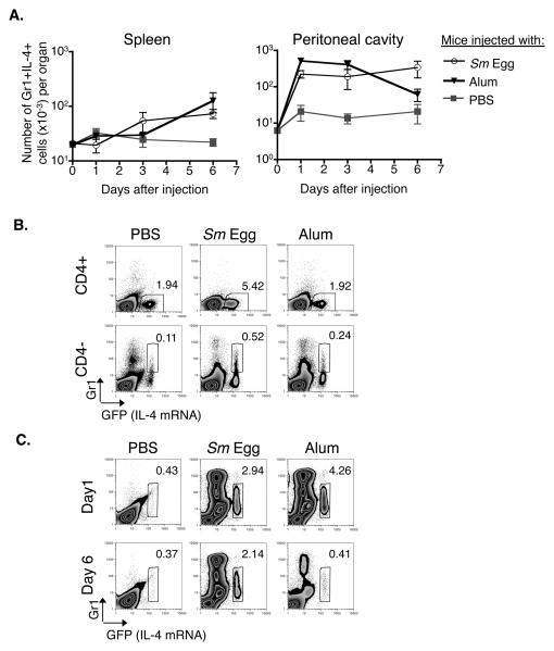
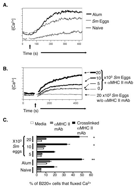
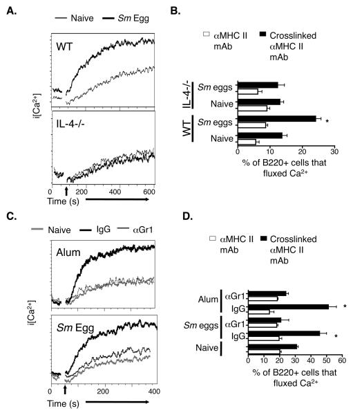
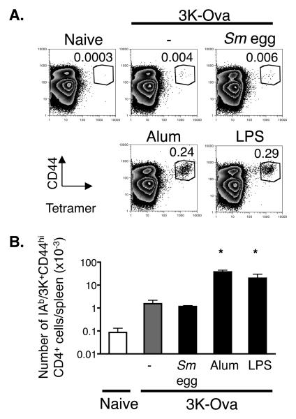
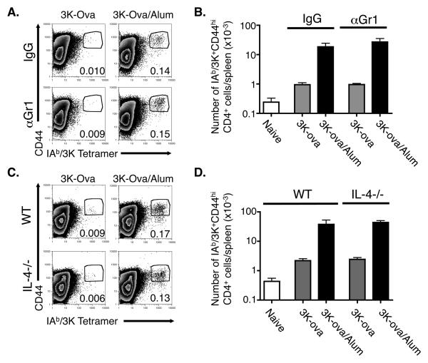
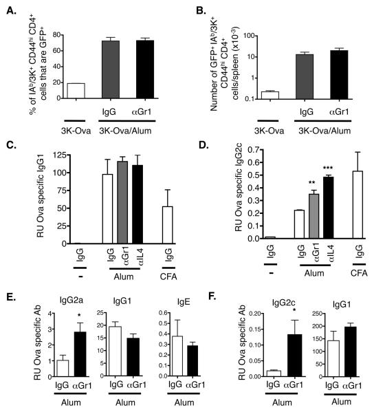
Alum is used as a vaccine adjuvant and induces T(h)2 responses and T(h)2-driven antibody isotype production against co-injected antigens. Alum also promotes the appearance in the spleen of Gr1+IL-4+ innate cells that, via IL-4 production, induce MHC II-mediated signaling in B cells. To investigate whether these Gr1+ cells accumulate in the spleen in response to other T(h)2-inducing stimuli and to understand some of their functions, the effects of injection of alum and eggs from the helminth, Schistosoma mansoni, were compared. Like alum, schistosome eggs induced the appearance of Gr1+IL-4+ cells in spleen and promoted MHC II-mediated signaling in B cells. Unlike alum, however, schistosome eggs did not promote CD4 T cell responses against co-injected antigens, suggesting that the effects of alum or schistosome eggs on splenic B cells cannot by themselves explain the T cell adjuvant properties of alum. Accordingly, depletion of IL-4 or Gr1+ cells in alum-injected mice had no effect on the ability of alum to improve expansion of primary CD4 T cells. However, Gr1+ cells and IL-4 played some role in the effects of alum, since depletion of either resulted in antibody responses to antigen that included not only the normal T(h)2-driven isotypes, like IgG1, but also a T(h)1-driven isotype, IgG2c. These data suggest that alum affects the immune response in at least two ways: one, independent of Gr1+ cells and IL-4, that promotes CD4 T cell proliferation and another, via Gr1+IL-4+ cells, that participates in the polarization of the response.


Figures

Figure 1
Figure 1. Sm eggs, like alum, induce the appearance of Gr1+IL-4+ innate cells
A. 4Get, IL-4 indicator mice were injected i.p. with alum, Sm eggs or PBS. At the indicated times, cells were isolated from the spleens and peritoneal cavities of the animals and stained with antibodies against CD4, CD8, B220 and Gr1 (splenocytes) or F480, cKit and Gr1 (peritoneal cells). Shown are the means and standard errors of numbers of B220, CD4 and CD8 negative splenocytes or F480 and cKit negative peritoneal cells that were Gr1+ IL-4+ (GFP+) isolated from 3 identically treated animals per group. B. Expression of Gr1 and GFP (IL-4 mRNA) on CD4 negative or CD4 positive spleen cells 6 days after i.p. injection with alum, Sm eggs or PBS. Numbers on graphs indicate the percent of gated CD4− or CD4+ cells that fall within the gates on each individual FACS plot. C. FACS plots show Gr1 and GFP (IL-4 mRNA) expression on F4/80- cKit- peritoneal cells from mice injected 1 or 6 days previously with PBS, Sm eggs, or alum. Numbers on graphs indicate the percent of F4/80- cKit- cells that fell within the gate on each plot for the sample shown. For B and C, data are representative FACS plots from 3 individual mice per group. 40mg alum or 104 Sm eggs were used for injections in this experiment. Data are representative of 3 independent experiments.
Figure 2
Figure 2. MHC II mediated Ca2+ responses in B cells are enhanced after exposure to Th2 stimuli
A. Spleen cells were isolated from mice injected 6 days previously with nothing (grey histogram), 104 Sm eggs (bold black histogram), or alum (fine black line histogram), and stained as described in Materials and Methods. MHC II molecules on the surface of the cells were crosslinked with biotinylated anti-MHC II followed by avidin. The time of addition of avidin is indicated on the Figure by the arrow. Shown are the changes in intracellular Ca2+ levels in B220+ cells. B. Mice were given the indicated numbers of Sm eggs. Six days later, spleen cells from the mice were stained and their MHC II molecules crosslinked as described in Figure 1A. Shown are the changes in intracellular Ca2+ induced by the crosslinking. Data in A and B are representative of 3 mice per group in a single experiment. C. Shown are the percent of B220+ cells undergoing Ca2+ flux in the absence of any stimulation (white bars), after addition of anti-MHC II mAb but before avidin mediated crosslinking (grey bars) and during the peak of the response after addition of avidin (solid bars) in mice injected 6 days previously with Sm egg, alum or PBS. Means +/− standard errors for values from 3 mice are shown and asterisks indicate results that differed from those in naïve mice with p<0.05 (*) or p<0.01 (**). Data in this Figure are from a representative experiment of 3.
Figure 3
Figure 3. The effects of Sm egg or alum on B cells are dependent upon IL-4 and Gr1 expressing cells
A. Wild type (WT) and IL-4−/− mice were injected i.p. with nothing (fine black line) or Sm eggs (bold black lines). Six days later, spleen cells were isolated and stained and MHC II crosslinked as described in Figure 2. Shown are the changes in intracellular Ca2+ levels in B220+ cells after MHC II crosslinking. B. Wild type or IL-4−/− mice were injected with nothing or Sm eggs and analyzed six days later as described in Figure 3A. Graphs show the percentage of splenic B220+ cells from either naïve, or Sm egg injected WT or IL-4−/− mice that that fluxed Ca2+ before (white bars) and during the peak (black bars) of the response to avidin mediated crosslinking of their MHC II molecules. C. Mice were injected i.p. nothing (grey lines), alum or Sm eggs (black lines). Alum or Sm egg injected mice were treated with either control rat IgG (bold black lines) or anti-Gr1 mAb (fine black lines). Six days after alum or Sm egg injection, spleen cells were isolated and their MHC II molecules crosslinked as described in Figure 2. Shown are the changes in intracellular Ca2+ levels in B220+ cells after MHC II crosslinking. D. Mice were injected with nothing, Sm eggs or alum, later with control rat IgG or anti-Gr1 and their spleen cells analyzed six days after injection of Sm eggs or alum as described in Figure 3C. Shown are the means +/− standard errors of the percentages of B220+ cells that had increased levels of intracellular Ca2+ before (white bars) or after (black bars) crosslinking of their MHC II proteins. Asterisks indicate a group with a percentage of cells fluxing Ca2+ after MHC II crosslinking that was significantly different from that observed in cells from naïve mice (p<0.05). Results in A and C are representative of two individual experiments and data in B and D are from a representative experiment of three.
Figure 4
Figure 4. Alum but not Sm eggs promotes enhanced CD4 responses to coinjected antigen
A. B6 mice were injected with 3K-Ova protein combined with either nothing (−), Sm eggs, alum, or 7μg LPS and splenocytes were stained with antibodies against B220, F480, CD44, CD4 and IAb/3K tetramers nine days later. Plots show staining with anti-CD44 and IAb/3K tetramers on CD4 positive, F4/80- B220- spleen cells. Numbers on graphs indicate the mean percentages of CD4 cells that were CD44hi IAb/3K tetramer positive for the sample shown. Data shown are representative of three identically treated mice in each group. Cells that stained as CD44hi and with IAb/3K tetramer in naïve mice illustrate the level of background staining in these experiments. B. Shown are the mean +/− standard errors of the numbers of IAb/3K specific CD44hi CD4 T cells detected per spleen for three mice per group. Asterisks indicate samples in which the numbers of IAb/3K specific T cells that were detected were significantly different from the numbers detected in mice injected with 3K-Ova alone (p<0.05). Data are from a representative experiment of three.
Figure 5
Figure 5. The ability of alum to enhance expansion of antigen specific CD4 cells is independent of Gr1 expressing cells and IL-4
A,B. C57BL/6 mice were injected with 3K-Ova either alone or adsorbed to alum and treated with either rat IgG or anti-Gr1 as described in Materials and Methods. Nine days later splenocytes were stained as in Figure 4. A. The FACS plots show the staining of F4/80- B220- CD4+ cells with anti-CD44 and IAb/3K tetramer. Numbers indicate the percentage of F4/80- B220- CD4+ cells that were CD44hi and IAb/3K tetramer positive and are representative of three individual samples. B. Shown are the average +/− standard error of the numbers of CD44hi IAb/3K-staining CD4+ cells/spleen of mice immunized as indicated. Results are representative of three independent experiments. C.D. Wild type or IL-4−/− mice were injected with 3K-Ova with or without alum. Nine days later their spleen cells were isolated and analyzed as in A, B. Results are representative of two separate experiments.
Figure 6
Figure 6. Gr1+ cells and IL-4 inhibit production of Th1-induced antibody isotypes
A, B. 4Get mice were injected with 3K-Ova with or without alum and were treated with either control rat IgG or anti-Gr1 mAb as described in Materials and Methods. Splenocytes were stained for the presence of IAb/3K specific CD4 T cells and gates were drawn around CD44hi IAb/3K+ CD4 cells as in Figs 4 and 5. A. Shown are the average +/−standard errors of the percentages of IAb/3K+ CD44hi CD4+ spleen cells that expressed GFP (IL-4 mRNA). The results are from three identically treated mice/group. B. Shown are the average numbers +/− standard errors of GFP+ IAb/3K+ CD44hi CD4+ cells/spleen from mice immunized as indicated. The results are from three identically treated mice/group. C, D. B6 mice were injected with Ova with or without alum or CFA. Mice were treated with IgG (white bars), anti-Gr1 (grey bars) or anti-IL-4 (black bars) on days one, three and five after primary immunization. On day nine mice were injected with Ova alone and on day twelve their sera were tested for Ova specific antibodies by ELISA as described in Materials and Methods. The results represent the mean relative units +/− standard error of Ova specific antibody detected from three individual mice per group. Asterisks indicate groups in which the amount of antibody detected was significantly different from that in similarly immunized mice that received control IgG. A single asterisk indicates p<0.05, double asterisk p<0.01, and triple asterisk indicates p<0.001. Results are representative of two independent experiments. E, F. Balb/c mice (E) or B6 mice (F) were injected with either alum (E) or Alhydrogel (F) adsorbed Ova and were treated with IgG (white bars) or αGr1 (black bars) antibody as above. On day 10, mice were boosted with alum adsorbed Ova and sera were tested for Ova specific isotypes 5 days later. Results are representative of 3 (E) and 2 (F) individual experiments.

Similar articles

Cited by

References

    1. Wille-Reece U, Flynn BJ, Lore K, Koup RA, Miles AP, Saul A, Kedl RM, Mattapallil JJ, Weiss WR, Roederer M, Seder RA. Toll-like receptor agonists influence the magnitude and quality of memory T cell responses after prime-boost immunization in nonhuman primates. J Exp Med. 2006;203:1249–58. - PMC - PubMed
    1. Kaisho T, Akira S. Toll-like receptors as adjuvant receptors. Biochim Biophys Acta. 2002;1589:1–13. - PubMed
    1. Medzhitov R, Janeway CA., Jr. Decoding the patterns of self and nonself by the innate immune system. Science. 2002;296:298–300. - PubMed
    1. Kawai T, Akira S. Pathogen recognition with Toll-like receptors. Curr Opin Immunol. 2005;17:338–44. - PubMed
    1. Hoebe K, Janssen E, Beutler B. The interface between innate and adaptive immunity. Nat Immunol. 2004;5:971–4. - PubMed

Publication types

MeSH terms