. 2008 May;7(5):791-9.
doi: 10.1128/EC.00009-08. Epub 2008 Mar 14.

MADS-box transcription factor mig1 is required for infectious growth in Magnaporthe grisea

Affiliations

MADS-box transcription factor mig1 is required for infectious growth in Magnaporthe grisea

Rahim Mehrabi et al. Eukaryot Cell. 2008 May.

Abstract

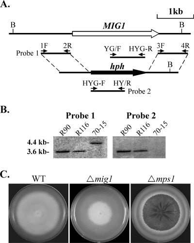
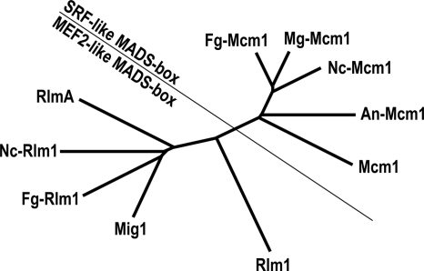
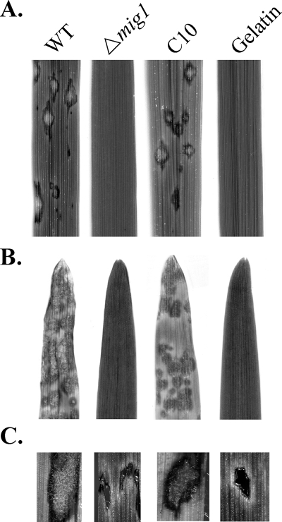
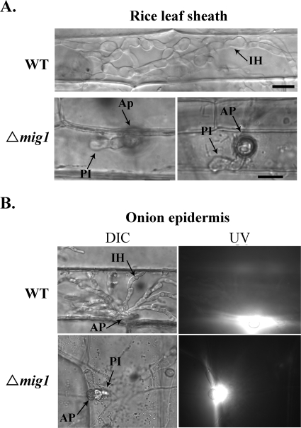
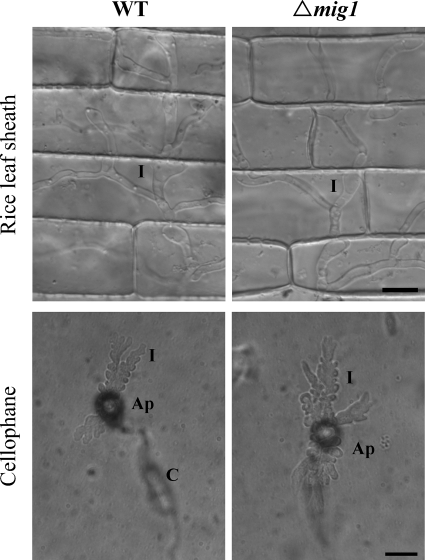
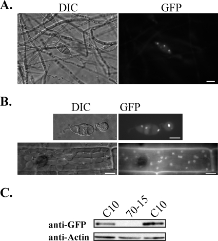
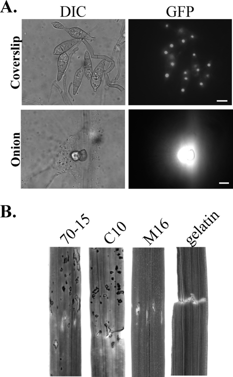
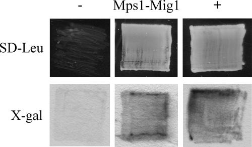
Magnaporthe grisea is a model fungus for studying fungus-plant interactions. Two mitogen-activated protein (MAP) kinase genes, PMK1 and MPS1, have been implicated in regulating plant infection processes in M. grisea. However, transcription factors activated by these MAP kinases are not well studied. In this study we functionally characterized the MIG1 gene that encodes a MADS-box transcription factor homologous to Saccharomyces cerevisiae Rlm1. In yeast two-hybrid assays, MIG1 interacts with MPS1, suggesting that MIG1 may function downstream from the MPS1 pathway. The mig1 deletion mutant had a normal growth rate and formed melanized appressoria, but it was nonpathogenic and failed to infect rice leaves through wounds. Appressoria formed by the mig1 mutant developed penetration pegs and primary infectious hyphae, but further differentiation of the secondary infectious hyphae inside live plant cells was blocked. However, the mig1 mutant formed infectious hypha-like structures in heat-killed plant cells or cellophane membranes. In transformants expressing the MIG1-GFP fusion, green fluorescent protein (GFP) signals were not detectable in vegetative hyphae and conidiophores. Mig1-GFP was localized to nuclei in conidia, appressoria, and infectious hyphae. Deletion of the MADS box had no effect on the expression and localization of the MIG1-GFP fusion but eliminated its ability to complement the mig1 mutant. These results suggest that MIG1 may be required for overcoming plant defense responses and the differentiation of secondary infectious hyphae in live plant cells. The MADS-box domain is essential for the function of MIG1 but dispensable for its nuclear localization, which may be associated with the activation of MIG1 by MPS1 during conidiation and plant infection.


Figures

FIG. 1.
FIG. 1.
Generation of mig1 deletion mutants. (A) The MIG1 gene replacement construct was generated by the split-marker approach. The small arrows mark the positions and directions of primers used in the PCRs. (B) Southern blots of BglII-digested genomic DNAs of mig1 deletion mutants (R90 and R116) and 70-15 were hybridized with probe 1 (left) and probe 2 (right). Probe 1 hybridized to a 4.4-kb fragment in 70-15 and a 3.6-kb fragment in R90 and R116. Probe 2 detected a 3.6-kb band in R90 and R116 but not in 70-15. (C) CM cultures of the wild-type (WT) strain and the mig1 and mps1 deletion mutants. The mig1 mutant was reduced in aerial hyphal growth and conidiation but had no defect in growth rate and cell wall integrity. Colony autolysis was observed only in the mps1 mutant.
FIG. 2.
FIG. 2.
The unrooted neighbor joining tree of the following putative MADS-box transcription factors from selected ascomycetes: Mcm1 (CAA88409) and Rlm1 (AAB68210) from Saccharomyces cerevisiae, MgMcm1 (EAA47530) and Mig1 (EU164776) from Magnaporthe grisea, NcMcm1 (EAA36453) and NcRlm1 (EAA35381) from Neurospora crassa, FgMcm1 (EAA76082) and FgRlm1 (EAA70796) from Fusarium graminearum, and AnMcm1 (EAA63555) and RlmA (EAA60098) from Aspergillus nidulans. SRF, serum response factor; MEF2, myocyte enhancer factor 2.
FIG. 3.
FIG. 3.
Infection assays with the wild-type strain (WT), the mig1 mutant R116 (Δmig1), and the complementation transformant of R116 (C10). Inoculation with 0.25% gelatin was used as the control. (A) Spray inoculation with 2-week-old rice seedlings. (B) Spray inoculation with 8-day-old barley seedlings. (C) Rice leaf segments containing the wound sites from injection-inoculation assays were surface sterilized and incubated on water agar for 2 days. Hyphal growth and conidiation were not observed on leaves inoculated with the mig1 mutant.
FIG. 4.
FIG. 4.
Penetration assays with the wild-type strain (WT) and the mig1 mutant (Δmig1). (A) Bulbous secondary infectious hyphae were observed only in rice leaf sheath epidermal cells penetrated by the wild-type strain. Some appressoria (about 3%) of the mig1 mutant were able to develop short primary infectious hyphae in plant cells. (B) The mig1 mutant elicited autofluorescence and papilla formation in onion epidermal cells but failed to develop secondary infectious hyphae. Images on the left and right were the same field examined under differential interference contrast (DIC) and epifluorescence microscopy (UV). Ap, appressorium; PI, primary infectious hyphae; IH, secondary infectious hyphae.
FIG. 5.
FIG. 5.
Penetration assays with heat-killed rice leaf sheaths and cellophane membranes. Both the wild-type strain (WT) and the mig1 mutant R116 (Δmig1) penetrated and developed infectious hypha-like structures in heat-killed rice leaf sheath epidermal cells of rice and cellophane membranes. Ap, appressorium; C, conidium; I, infectious hypha-like structures. Bars = 10 μm.
FIG. 6.
FIG. 6.
Expression and localization of Mig1-GFP in transformant C10. (A) GFP signals were observed in the nuclei of conidia but not in aerial hyphae or conidiophores harvested from 9-day-old oatmeal cultures. The same field was examined under differential interference contrast (DIC) and epifluorescence microscopy (GFP). Bar = 10 μm. (B) GFP signals were preferentially localized to the nuclei in appressoria formed on glass coverslips and infectious hyphae produced in rice leaf sheath epidermal cells. Bars = 10 μm. (C) Western blot analysis with proteins isolated from vegetative hyphae of the wild-type strain 70-15 and transformant C10. An 86-kDa band of the expected size of Mig1-GFP fusion was detected with an anti-GFP antibody.
FIG. 7.
FIG. 7.
The MADS box is essential for the function of Mig1. (A) Deletion of the MADS box had no effect on the expression and localization of the MIG1-GFP fusion. Fluorescent signals were observed in the nuclei of conidial cells but not in aerial hyphae of MIG1ΔMADS-GFP transformant M16 (top panels). Appressoria formed by transformant M16 elicited papilla formation and autofluorescence in underlying plant cells but failed to develop infectious hyphae in onion epidermal cells (bottom panels). Bars = 10 μm. (B) Rice leaves wound-inoculated with the wild-type (70-15), complementation transformant C10, MIG1ΔMADS-GFP transformant M16, and 0.25% gelatin.
FIG. 8.
FIG. 8.
Yeast two-hybrid assays. Yeast transformants containing the bait and prey constructs of MIG1 and MPS1 were able to grow on SD-Leu plates and express β-galactosidase activities when galactose was used as the carbon source. Transformants expressing pRFHM1 and pSH18-34 or pSH17-4 and pSH18-34 were used as the negative (−) and positive (+) controls, respectively. Growth on SD-Leu plates and β-galactosidase activities were examined after incubation for 48 h. X-gal (5-bromo-4-chloro-3-indolyl-β-d-galactopyranoside) was used as the substrate to assay galactosidase activities.

References

    1. Bourett, T. M., and R. J. Howard. 1990. In vitro development of penetration structures in the rice blast fungus Magnaporthe grisea. Can. J. Bot. 68329-342.
    1. Bruno, K. S., F. Tenjo, L. Li, J. E. Hamer, and J.-R. Xu. 2004. Cellular localization and role of kinase activity of PMK1 in Magnaporthe grisea. Eukaryot. Cell 31525-1532. - PMC - PubMed
    1. Catlett, N. L., B. Lee, O. C. Yoder, and B. G. Turgeon. 2003. Split-marker recombination for efficient targeted deletion of fungal genes. Fungal Genet. Newsl. 509-11.
    1. Chao, C. C. T., and A. H. Ellingboe. 1991. Selection for mating competence in Magnaporthe grisea pathogenic to rice. Can. J. Bot. 692130-2134.
    1. Choi, W. B., and R. A. Dean. 1997. The adenylate cyclase gene MAC1 of Magnaporthe grisea controls appressorium formation and other aspects of growth and development. Plant Cell 91973-1983. - PMC - PubMed

Publication types

MeSH terms

LinkOut - more resources