Comparative Study
. 2008 Aug;147(4):1590-602.
doi: 10.1104/pp.108.116863. Epub 2008 Mar 14.

Comparison of the dynamics and functional redundancy of the Arabidopsis dynamin-related isoforms DRP1A and DRP1C during plant development

Affiliations
Comparative Study

Comparison of the dynamics and functional redundancy of the Arabidopsis dynamin-related isoforms DRP1A and DRP1C during plant development

Catherine A Konopka et al. Plant Physiol. 2008 Aug.

Abstract

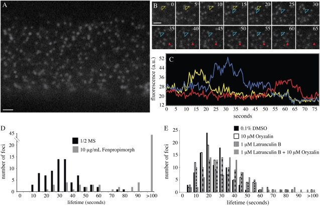
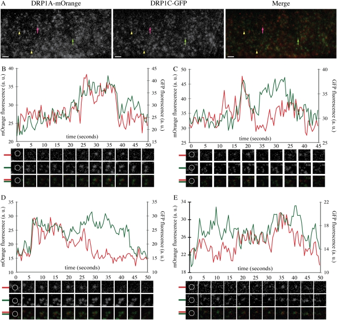
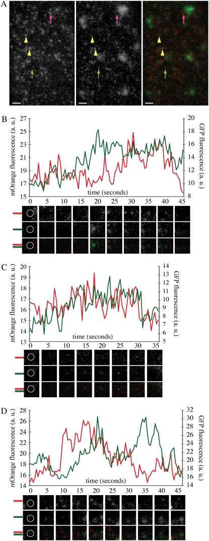
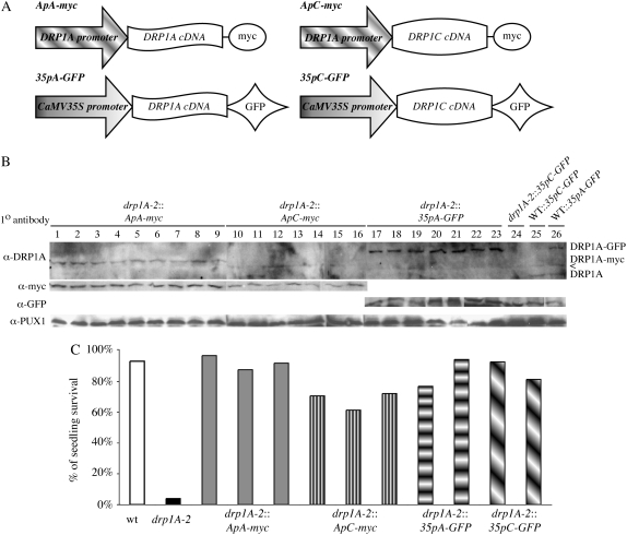
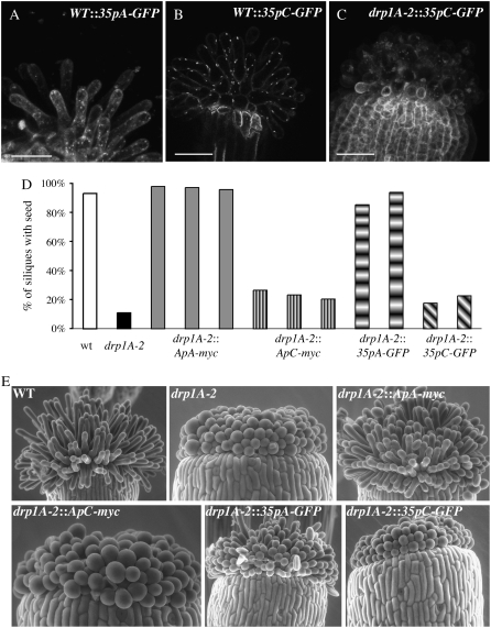
Members of the Arabidopsis (Arabidopsis thaliana) DYNAMIN-RELATED PROTEIN1 (DRP1) family are required for cytokinesis and cell expansion. Two isoforms, DRP1A and DRP1C, are required for plasma membrane maintenance during stigmatic papillae expansion and pollen development, respectively. It is unknown whether the DRP1s function interchangeably or if they have distinct roles during cell division and expansion. DRP1C was previously shown to form dynamic foci in the cell cortex, which colocalize with part of the clathrin endocytic machinery in plants. DRP1A localizes to the plasma membrane, but its cortical organization and dynamics have not been determined. Using dual color labeling with live cell imaging techniques, we showed that DRP1A also forms discreet dynamic foci in the epidermal cell cortex. Although the foci overlap with those formed by DRP1C and clathrin light chain, there are clear differences in behavior and response to pharmacological inhibitors between DRP1A and DRP1C foci. Possible functional or regulatory differences between DRP1A and DRP1C were supported by the failure of DRP1C to functionally compensate for the absence of DRP1A. Our studies indicated that the DRP1 isoforms function or are regulated differently during cell expansion.


Figures

Figure 1.
Figure 1.
Phylogenetic tree of DRPs in Arabidopsis, rice, and Medicago. ClustalW alignment of the entire primary amino acid sequence of DRPs identified in BLAST searches from the published Arabidopsis, Oryza, and Medicago genomes and human dynamin 1, yeast Vps1, and soybean phragmoplastin were used to generate the phylogenetic tree. The scale represents to the number of nucleotide substitution events based on amino acid differences.
Figure 2.
Figure 2.
Analysis of DRP1A-GFP focus dynamics at the cell cortex. A, Epidermal root cell in the elongation zone expressing DRP1A-GFP imaged with VAEM. B, Image montage taken from time lapse sequence of the epidermal root cell shown in A. Numbers in top right corners indicate time elapsed from first image in seconds. The focus indicated by the open arrowhead changed position during its lifetime in the cell cortex (first position, yellow arrowhead; second position, blue arrowhead). The focus indicated by the solid red arrowhead did not change position. C, Intensity profiles of the foci indicated in B. The mobile focus (open arrowhead) is indicated by both yellow (first position) and blue (second position) lines. D, Lifetime distribution of DRP1A-GFP foci in cells from plants grown on one-half-strength Murashige and Skoog (1/2 MS) with no drug (black bars) and from plants grown in the presence of 10 μg/mL fenpropimorph (gray bars). E, Lifetime analysis of DRP1A-GFP from plants treated for 20 min with 10 μm oryzalin (white bars), 1 μm latB (striped bars), concurrently with 10 μm oryzalin + 1 μm latB (gray bars), or mock treated with 0.1% DMSO (black bars). Scale bars = 1 μm.
Figure 3.
Figure 3.
DRP1A-mOrange foci colocalize with DRP1C-GFP foci in the cell cortex. A, Epidermal root cell in the expansion zone expressing DRP1A-mOrange and DRP1C-GFP imaged with VAEM with filter set for simultaneous GFP and mOrange fluorescence capture (see “Materials and Methods”). DRP1A-mOrange and DRP1C-GFP foci are present without the other DRP1 (DRP1A, pink arrow; DRP1C, green arrow) and also colocalize (yellow arrowheads). B to E, Intensity profiles of GFP (green) and mOrange (red) fluorescence from foci that had overlapping fluorescence of DRP1C-GFP and DRP1A-mOrange. Corresponding mOrange fluorescence (top), GFP fluorescence (middle), and merged (bottom) images are below each time (in seconds) indicated in the graph. Montage images are 1.2 × 1.2 μm. Yellow circles in the first frames indicate measured regions for fluorescence intensity. Bars = 1 μm.
Figure 4.
Figure 4.
DRP1A-mOrange foci colocalize with CLC-GFP foci in the cell cortex. A, Epidermal root cell in the expansion zone expressing DRP1A-mOrange and CLC-GFP imaged with VAEM with filter set for simultaneous GFP and mOrange fluorescence capture (see “Materials and Methods”). DRP1A-mOrange and CLC-GFP foci are present independently (DRP1A, pink arrow; CLC, green arrow) and also colocalize (yellow arrowheads). B to D, Intensity profiles of GFP (green) and mOrange (red) fluorescence from foci that had overlapping fluorescence of CLC-GFP and DRP1A-mOrange. Corresponding mOrange fluorescence (top), GFP fluorescence (middle), and merged (bottom) images are below each time (in seconds) indicated in the graph. Montage images are 1.2 × 1.2 μm. White circles in the first frames indicate measured regions for fluorescence intensity. Bars = 1 μm.
Figure 5.
Figure 5.
Exogenous expression of DRP1C rescues drp1A-2 seedling lethality. A, Schematic of the four constructs used for complementation analysis. B, Immunoblot of total protein extracts from drp1A-2 (lanes 1–24) or wild-type (lanes 25 and 26) seedlings expressing ApA-myc (lanes 1–9), ApC-myc (lanes 10–16), 35pA-GFP (lanes 17–23, 26), or 35pC-GFP (lane 24). The top blot was blotted with anti-DRP1A specific antibodies, the middle blot with anti-myc (left) or anti-GFP (right) antibodies, and the bottom blot with anti-PUX1 antibodies (loading control). All lines express the transgene approximately equally well. The bands in the anti-DRP1A blot were DRP1A-GFP (top), DRP1A-myc (middle), and native, untagged DRP1A (bottom). A cross-reactive band is indicated by <. C, Histogram indicating the percentage of seedlings of individual lines (with the genotype indicated) that survived and developed a second set of true leaves on one-half-strength Murashige and Skoog, 0.6% agar without Suc.
Figure 6.
Figure 6.
Expression of DRP1C cannot rescue the expansion defect of drp1A-2 stigmatic papillae. A to C, Maximal Z projections (A and C) or a single confocal section (B) of stigmatic papillae from first stage 13 flower from plants with genotypes indicated. C, Histogram indicating the percent of siliques in individual lines with the genotype indicated that had at least one seed. Siliques from four to six plants from each line were evaluated for seed content. D, Environmental scanning electron microscopy images of stigmatic papillae from the first open flower from plants with the genotypes indicated. Bars = 50 μm.

References

    1. Arabidopsis Genome Initiative (2000) Analysis of the genome sequence of the flowering plant Arabidopsis thaliana. Nature 408 796–815 - PubMed
    1. Banbury DN, Oakley JD, Sessions RB, Banting G (2003) Tyrphostin A23 inhibits internalization of the transferrin receptor by perturbing the interaction between tyrosine motifs and the medium chain subunit of the AP-2 adaptor complex. J Biol Chem 278 12022–12028 - PubMed
    1. Baskin TI, Wilson JE, Cork A, Williamson RE (1994) Morphology and microtubule organization in Arabidopsis roots exposed to oryzalin or taxol. Plant Cell Physiol 35 935–942 - PubMed
    1. Clough SJ, Bent AF (1998) Floral dip: a simplified method for Agrobacterium-mediated transformation of Arabidopsis thaliana. Plant J 16 735–743 - PubMed
    1. Conner SD, Schmid SL (2003) Regulated portals of entry into the cell. Nature 422 37–44 - PubMed

Publication types

MeSH terms