Comparative Study
. 2008 Apr;9(4):356-62.
doi: 10.1038/embor.2008.28. Epub 2008 Mar 14.

Clathrin-independent endocytosis used by the IL-2 receptor is regulated by Rac1, Pak1 and Pak2

Affiliations
Comparative Study

Clathrin-independent endocytosis used by the IL-2 receptor is regulated by Rac1, Pak1 and Pak2

Alexandre Grassart et al. EMBO Rep. 2008 Apr.

Abstract

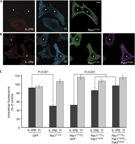
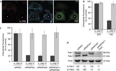
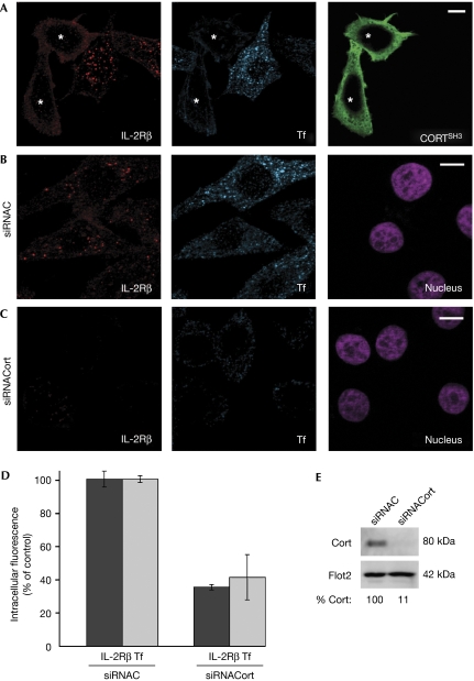
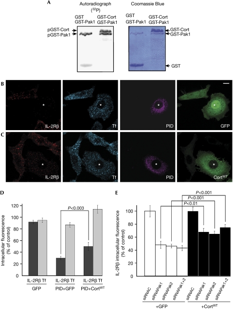
There are several endocytic pathways, which are either dependent on or independent of clathrin. This study focuses on a poorly characterized mechanism-clathrin- and caveolae-independent endocytosis-used by the interleukin-2 receptor beta (IL-2R beta). We address the question of its regulation in comparison with the clathrin-dependent pathway. First, we show that Ras-related C3 botulinum toxin substrate 1 (Rac1) is specifically required for IL-2R beta entry, and we identify p21-activated kinases (Paks) as downstream targets. By RNA interference, we show that Pak1 and Pak2 are both necessary for IL-2R beta uptake, in contrast to the clathrin-dependent route. We observe that cortactin, a partner of actin and dynamin-two essential endocytic factors-is required for IL-2R beta uptake. Furthermore, we find that cortactin acts downstream from Paks, suggesting control of its function by these kinases. Thus, we describe a cascade composed of Rac1, Paks and cortactin specifically regulating IL-2R beta internalization. This study indicates Paks as the first specific regulators of the clathrin-independent endocytosis pathway.


Conflict of interest statement

The authors declare that they have no conflict of interest.

Figures

Figure 1
Figure 1
A Rac1 dominant-negative mutant inhibits specifically IL-2Rβ endocytosis and can be counteracted by an active form of Pak1. (A) Endocytosis of IL-2Rβ (red) and Tf (blue) were examined by immunofluorescence in Hep2β cells transfected with a dominant-negative mutant of Rac1, myc-Rac1T17N (green). The cells were incubated for 15 min at 37°C in the presence of Cy3-coupled IL-2Rβ antibody and Alexa Fluor 647-coupled Tf. Cells were then fixed, permeabilized and reacted with myc antibody and the corresponding secondary antibody. A medial section is shown; asterisks indicate the cells expressing myc-Rac1T17N. (B) Hep2β cells were transfected with myc-Rac1T17N and a constitutively active form of Pak1, HA-Pak1T423E. Endocytosis of IL-2Rβ and Tf were followed as described in (A). (C) Quantification of (A), (B) and Hep2β cells transfected with myc-Rac1T17N and GFP, as well as Hep2β cells transfected with myc-Rac1T17N, HA-Pak1T423E and a constitutively active form of Pak2, GFP-Pak2T423E. To quantify endocytosis, the intracellular fluorescence intensity was measured with Metamorph software (mean±s.e.; n≈100 cells from three independent experiments). The results are expressed as a percentage of the intracellular fluorescence intensity of non-transfected cells. GFP, green fluorescent protein; HA, haemagglutinin; IL-2Rβ, interleukin-2 receptor β; Pak, p21-activated kinase; Rac1, Ras-related C3 botulinum toxin substrate 1; Tf, transferrin.
Figure 2
Figure 2
Pak1 and Pak2 are necessary for IL-2Rβ entry but not for transferrin uptake. (A) Endocytosis of IL-2Rβ (red) and Tf (blue) in cells transfected with the Pak1 inhibitory domain, myc-PID (green). Endocytosis and immunofluorescence were carried out as described in Fig 1A (asterisks indicate the cells expressing myc-PID). (B) Quantification of (A) and of Hep2β cells transfected with GFP as a control was carried out as described in Fig 1C. (C) Quantification of intracellular IL-2Rβ and Tf in Pak1- and/or Pak2-knockdown cells. Hep2β cells were transfected with small interfering RNA (siRNA) against Pak1 (siRNAPak1), Pak2 (siRNAPak2) or against an irrelevant protein (siRNAC). Endocytosis and quantification were carried out for 200 cells. The results are expressed as a percentage of the intracellular fluorescence intensity of control cells (siRNAC). (D) Western blots of siRNA-transfected cells were probed with antibodies against Pak1, Pak2 or against flotillin 2 (Flot2) as a control; quantification by Storm FluoroImager. GFP, green fluorescent protein; IL-2Rβ, interleukin-2 receptor β; Pak, p21-activated kinase; PID, Pak inhibitory domain; Rac1, Ras-related C3 botulinum toxin substrate 1; Tf, transferrin.
Figure 3
Figure 3
Cortactin is necessary for IL-2Rβ and for transferrin endocytosis. (A) Hep2β cells transfected with a dominant-negative mutant of cortactin, Flag-CortSH3 (green). (B) Cells treated with control small interfering RNA (siRNAC). (C) Cells treated with siRNA against cortactin (siRNACort). (AC) Endocytosis and immunofluorescence were carried out as described in Fig 1A (asterisks indicate the cells expressing CortSH3). Nuclei were stained with Hoechst (purple). (D) Quantification of IL-2Rβ and Tf endocytosis was carried out as described in Fig 2C. (E) Western blots of siRNA-transfected cells were probed with antibodies against cortactin or flotillin 2 (Flot2) as a control; quantification by Storm FluoroImager. IL-2Rβ, interleukin-2 receptor β; Pak, p21-activated kinase; Rac1, Ras-related C3 botulinum toxin substrate 1; Tf, transferrin.
Figure 4
Figure 4
Cortactin acts downstream of Paks in IL-2Rβ endocytosis. (A) In vitro phosphorylation of cortactin by Pak1. Purified GST-Pak1 (1 μg) was incubated with 5 μg of purified GST-cortactin (GST-Cort) or GST and 5 μCi γ[32P]ATP. Autoradiography (left), Coomassie blue gel (right). (B) Hep2β cells were transfected with myc-PID and GFP. (C) Hep2β cells were transfected with myc-PID and GFP-cortactin wild-type (CortWT). Cells were treated and analysed as described in Fig 1 (asterisks indicate the cells coexpressing the constructs). (D) Quantification of the endocytosis results was carried out as described in Fig 1C. (E) Hep2β cells were transfected with small interfering RNA (siRNA) against Pak1 (siRNAPak1) and/or Pak2 (siRNAPak2) or against an irrelevant protein (siRNAC) and transfected either with GFP-cortactin or GFP. Quantification of the endocytosis results was carried out as described in Fig 1C (mean±s.e.; n=50 cells in two independent experiments). GFP, green fluorescent protein; GST, glutathione-S-transferase; IL-2Rβ, interleukin-2 receptor β; Pak, p21-activated kinase; PID, Pak inhibitory domain; Rac1, Ras-related C3 botulinum toxin substrate 1; Tf, transferrin.

References

    1. Burridge K, Doughman R (2006) Front and back by Rho and Rac. Nat Cell Biol 8: 781–782 - PubMed
    1. Cao H, Orth JD, Chen J, Weller SG, Heuser JE, McNiven MA (2003) Cortactin is a component of clathrin-coated pits and participates in receptor-mediated endocytosis. Mol Cell Biol 23: 2162–2170 - PMC - PubMed
    1. Cheng ZJ, Singh RD, Sharma DK, Holicky EL, Hanada K, Marks DL, Pagano RE (2006) Distinct mechanisms of clathrin-independent endocytosis have unique sphingolipid requirements. Mol Biol Cell 17: 3197–3210 - PMC - PubMed
    1. Daly RJ (2004) Cortactin signalling and dynamic actin networks. Biochem J 382: 13–25 - PMC - PubMed
    1. Dharmawardhane S, Schurmann A, Sells MA, Chernoff J, Schmid SL, Bokoch GM (2000) Regulation of macropinocytosis by p21-activated kinase-1. Mol Biol Cell 11: 3341–3352 - PMC - PubMed

Publication types