

Comparative Study
doi: 10.1038/nn2073.
Epub 2008 Mar 16.
ALS-causing SOD1 mutants generate vascular changes prior to motor neuron degeneration
Affiliations
- PMID: 18344992
- PMCID: PMC2895310
- DOI: 10.1038/nn2073
Item in Clipboard
Comparative Study
ALS-causing SOD1 mutants generate vascular changes prior to motor neuron degeneration
Nat Neurosci.
2008 Apr.
Abstract
We report here that amyotrophic lateral sclerosis-linked superoxide dismutase 1 (SOD1) mutants with different biochemical characteristics disrupted the blood-spinal cord barrier in mice by reducing the levels of the tight junction proteins ZO-1, occludin and claudin-5 between endothelial cells. This resulted in microhemorrhages with release of neurotoxic hemoglobin-derived products, reductions in microcirculation and hypoperfusion. SOD1 mutant-mediated endothelial damage accumulated before motor neuron degeneration and the neurovascular inflammatory response occurred, indicating that it was a central contributor to disease initiation.
Figures
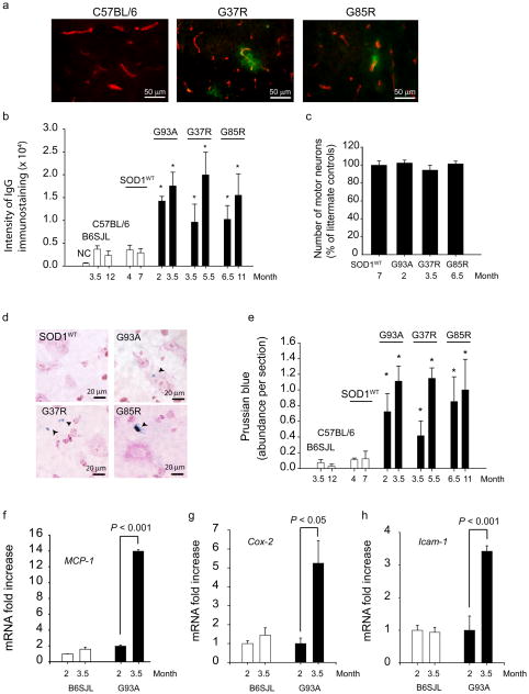
(a) Double immunostaining for IgG (green) and CNS endothelium (CD31, red) in the lumbar spinal cord of wild type mouse and 3.5 months old SOD1G37R and 6.5 months old SOD1G85R mutants. (b) Quantification of the IgG signal intensity in the lumbar spinal cord of different SOD1 mutants at varying ages compared to their respective controls and SOD1WT mice. NC, negative controls (omission of IgG antibody). (c) The number of motor neurons in the lumbar cord of different SOD1 mutants at an early stage of the disease process and in SOD1WT mice expressed as a percentage of total number of motor neurons in non-transgenic controls. (d) Hemosiderin extra-neuronal and neuronal deposition in the lumbar spinal cords in different SOD1 mutants, but not in SOD1WT mice. (e) Quantification of hemosiderin deposits in the lumbar spinal cord of different SOD1 mutants, controls and SOD1WT mice. (f–h) Quantitative RT-PCR analysis of mRNA transcripts for MCP-1, Cox-2 and Icam-1 in the lumbar spinal cord of SOD1G93A mice and controls at different age. Values are means ± s.e.m., n = 3–6 mice per group. *P < 0.05 compared to SOD1WT or non-transgenic mice. All procedures were according to the NIH guidelines and approved by the University of Rochester Committee on Animal Resources.
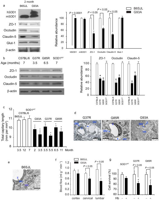
(a) Immunoblot analysis of human hSOD1, mouse mSOD1, ZO-1, occludin and claudin-5, and Glut-1 in spinal cord microvessels from 2 months old SOD1G93A mice compared to controls. Graph - band density for test-proteins relative to β-actin. (b) Immunoblot analysis of ZO-1, occludin and claudin-5 in the spinal cord capillaries from 7, 3.5 and 6.5 months old C57BL/6, mice, SOD1G37R and SOD1G85R mutants, respectively, and 7 months old SOD1WT mice. Graph - band density for test-proteins relative to β-actin. *P < 0.05, SOD1 mutants vs. controls or SOD1WT mice. (c) Total capillary length in the anterior horn of the lumbar spinal cord in SOD1 mutants (mm CD31-positive structures per mm2). *P < 0.05, SOD1 mutants vs. age matched controls. (d–e) Transmission electron microscopy (TEM) in 3.5 and 6.5 months old SOD1G37R and SOD1G85R mutants, respectively, shows edema (asterisk) between the capillary wall and motor neurons. Collapsed capillary lumen in SOD1G93A mice. E, endothelium; BM, basement membrane, MN, motor neuron. (e) TEM of B6SJL mouse shows normal neuronal-vascular contact and intact endothelium. (f) Blood flow through the spinal cord and frontal cortex in 8 weeks old SOD1G93A mice compared to controls. (g) Hemoglobin (Hb) toxicity to N2a cells transduced with SOD1G37R and SOD1G85R mutants and SOD1WT. #P < 0.05, SOD1G37R or SOD1G85R vs. SOD1WT. Mean ± s.e.m. a–c and f, n = 3–5 mice per group. g, n = 4 experiments per group.
Comment in
-
Implications of blood-brain barrier disruption in ALS.Amyotroph Lateral Scler. 2008 Dec;9(6):375-6. doi: 10.1080/17482960802160990. Amyotroph Lateral Scler. 2008. PMID: 18608097
References
-
- Boillée S, Vande Velde C, Cleveland DW. ALS: a disease of motor neurons and their nonneuronal neighbors. Neuron. 2006;52:39–59. - PubMed
-
- Boillée S, et al. Onset and progression in inherited ALS determined by motor neurons and microglia. Science. 2006;312:1389–1392. - PubMed
-
- Garbuzova-Davis S, et al. Ultrastructure of blood-brain barrier and blood-spinal cord barrier in SOD1 mice modeling ALS. Brain Res. 2007;1157:126–137. - PubMed
-
- Pun S, et al. Selective vulnerability and pruning of phasic motorneuron axons in motorneuron disease alleviated by CNTF. Nat Neurosci. 2006;9:408–419. - PubMed
Publication types
MeSH terms
Substances
Grants and funding
LinkOut - more resources
Full Text Sources
Other Literature Sources
Medical
Molecular Biology Databases
Miscellaneous
