. 2007 May;3(2):127-40.
doi: 10.1017/S1740925X07000440.

Added astroglia promote greater synapse density and higher activity in neuronal networks

Affiliations

Added astroglia promote greater synapse density and higher activity in neuronal networks

Michael D Boehler et al. Neuron Glia Biol. 2007 May.

Abstract

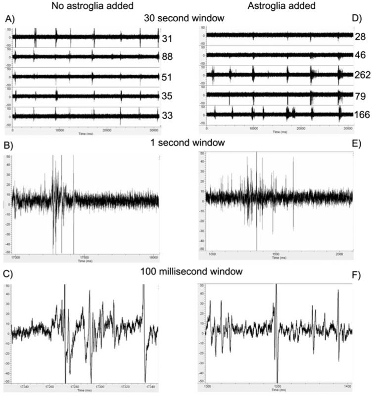
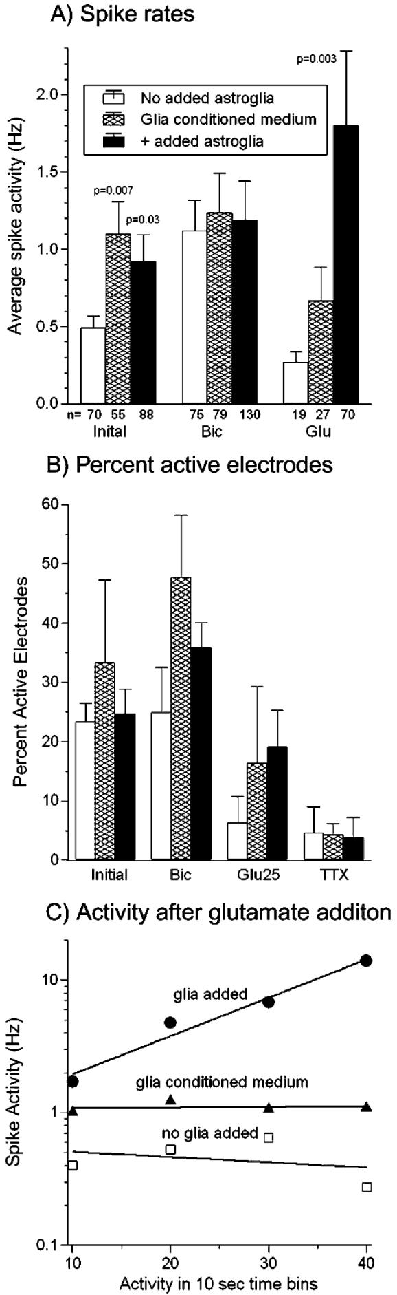
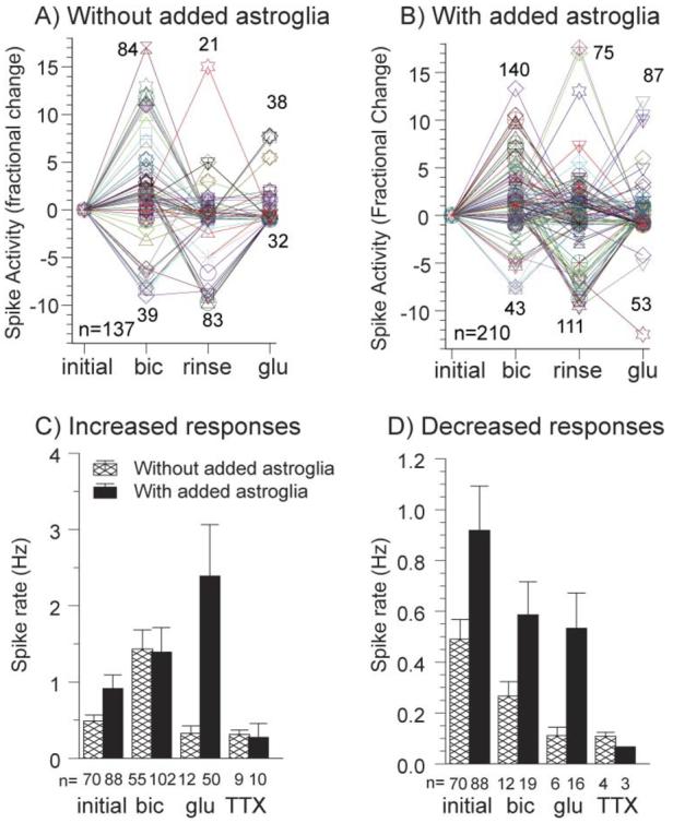
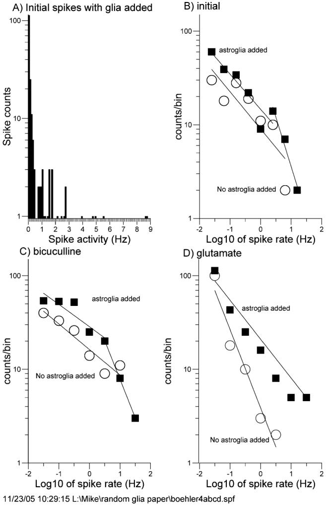
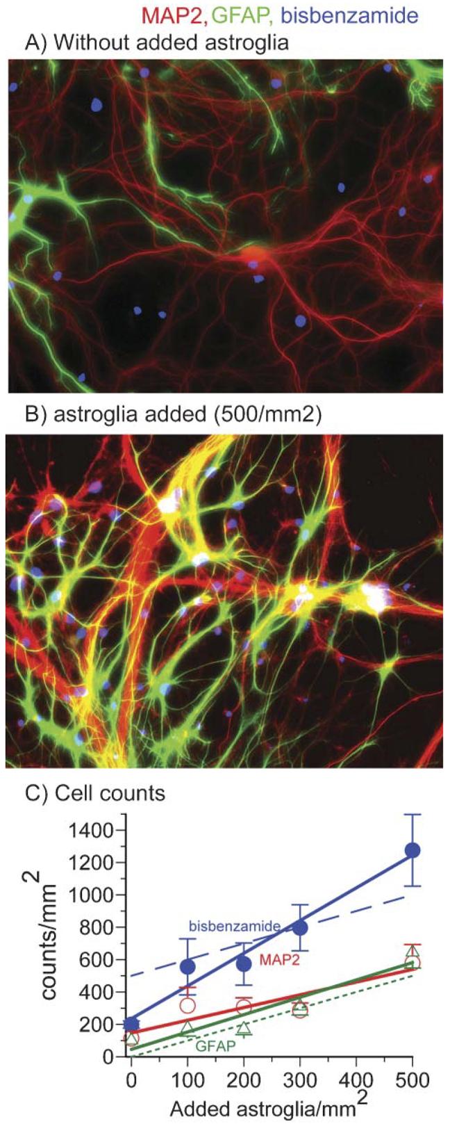
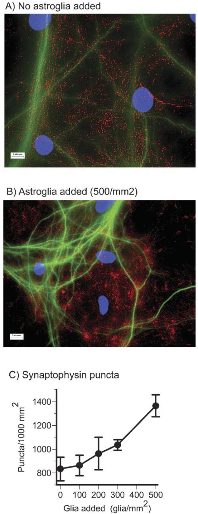
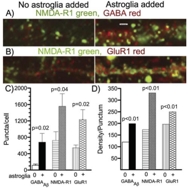
Astroglia are known to potentiate individual synapses, but their contribution to networks is unclear. Here we examined the effect of adding either astroglia or media conditioned by astroglia on entire networks of rat hippocampal neurons cultured on microelectrode arrays. Added astroglia increased spontaneous spike rates nearly two-fold and glutamate-stimulated spiking by six-fold, with desensitization eliminated for bath addition of 25 microM glutamate. Astrocyte-conditioned medium partly mimicked the effects of added astroglia. Bursting behavior was largely unaffected by added astroglia except with added glutamate. Addition of the GABA(A) receptor antagonist bicuculline also increased spike rates but with more subtle differences between networks without or with added astroglia. This indicates that networks without added astroglia were inhibited greatly. In all conditions, the log-log distribution of spike rates fit well to linear distributions over three orders of magnitude. Networks with added astroglia shifted consistently toward higher spike rates. Immunostaining for GFAP revealed a linear increase with added astroglia, which also increased neuronal survival. The increased spike rates with added astroglia correlated with a 1.7-fold increase in immunoreactive synaptophysin puncta, and increases of six-fold for GABA(Abeta), two-fold for NMDA-R1 and two-fold for Glu-R1 puncta, with receptor clustering that indicated synaptic scaling. Together, these results indicate that added astroglia increase the density of synapses and receptors, and facilitate higher spike rates for many elements in the network. These effects are reproduced by glia-conditioned media, with the exception of glutamate-mediated transmission.

Keywords: multielectrode array; neuron–glia cell culture; serum-free culture; synaptic scaling; synaptogenesis.


Figures

Fig. 1
Fig. 1. More action potentials are detected on multielectrode arrays to which astroglia were added
Action potential activity of cultures (22 days in vitro) without added astroglia (A,B,C) and with added astroglia (D,E,F). Traces from five electrodes for 30 sec of spike activity with added astroglia (D) have more activity and longer bursts than without added astroglia (A). Numbers indicate spike counts at +5 S.D. above noise. Spike waveforms and shape are similar between groups at 1 sec (B,E) and 100 msec (C,F).
Fig. 2
Fig. 2. Average spike rates increase with added astroglia and in response to glutamate with less desensitization
(A) Initial spike rates show a 1.9-fold mean increase for arrays with added astroglia and 2.0-fold increase with glia-conditioned medium compared to arrays without added astroglia. Bicuculline addition raised spike rates and the number of active channels (n), however, there was no difference in rate between networks with and without added astroglia or with glia-conditioned medium. Glutamate addition to cultures with astroglia produced a 6.7-fold mean increase in activity over arrays without added astroglia (P=0.003) and a 2.6-fold increase over glia-conditioned medium. P values show significant differences from no glia added condition by t-test. (B) Active electrodes per 60 electrode array tends to increase with added astroglia or glia-conditioned medium (letter-toe) (n = 300 electrodes) compared to no added astroglia. (C) Spike activity immediately following the addition of glutamate in 10-sec bins shows increased activity 10–40 sec after addition of glutamate in a culture with added astroglia (●, slope = 0.029, n=28–51 electrodes). A decrease or lower activity is seen over the same time period in a culture without added astroglia (□, slope = −0.004, n=2–8 electrodes) and in a culture with glia-conditioned medium (▲, slope=0.0004, n=14–34 electrodes). In all evaluations, electrodes with either 0 or 1 spike were not included in the averages. Cultures were 22–29 days in vitro.
Fig. 3
Fig. 3. Separation of increased from decreased spike rates on each electrode increases effect of added astroglia with glutamate to 7-fold
Fold increase or decrease in spike rate for individual electrodes after initial activity in cultures without added astroglia (A) and cultures with added astroglia (B). Changes are relative to the preceding condition with numbers of responding electrodes indicated near the peaks and numbers of electrodes active under any condition in the lower left. (C,D) Mean rates for cultures with added astroglia and without added astroglia for increased responses (C) and decreased responses (D) relative to initial activity. For increased responses, after glutamate addition, cultures with astroglia added have >7-fold higher activity than cultures without added astroglia (C) responses (P=0.003). For decreased responses after glutamate (D), cultures with added astroglia show 5-fold more activity (P=0.008). For the small fraction of decreased responses after bicuculline (D), cultures with astroglia added have 2-fold higher activity than cultures without added astroglia (P=0.03). Cultures were 22–29 DIV.
Fig. 4
Fig. 4. Log–log distribution analysis of spike rates shows increased spike rates for added astroglia
(A) Spike-frequency distribution shows a non-Gaussian peak with one tail. (B,C,D) Log–log distributions of networks without added astroglia (○) and with added astroglia (■) for initial condition (B), after bicuculline (C) and after glutamate (D). Parameters of regression-line fits are listed in Table 3. In all cases, cultures with added astroglia show a distribution shift to higher frequencies. Cultures were 22–29 DIV.
Fig. 5
Fig. 5. Added astroglia improves neuron survival.
(A,B) Immunostaining for astroglial GFAP (green) and neuronal MAP2 (red) after incubation for 22 days indicates more astroglia are present when (B) astroglia are added to the cultures than when (A) no astroglia were added. Nuclear staining with bisbenzamide appears blue. (C) Regression lines show the effect of adding increasing concentrations of astroglia on immunostaining of nuclei (bisbenzamide, ●), neurons (MAP2, ○) and astroglia (GFAP, △). Immunostaining by each increases as more astroglia are added. The dotted line is a theoretical result assuming no astroglia present when none were added. The dashed line is the theoretical total nuclei assuming all neurons survive and astroglia start at zero. Note that actual astroglial counts start at 100 after 22 days of culture not zero and bisbenzamide counts start at 200 after 22 days, not the 500 cells that were originally added.
Fig. 6
Fig. 6. Added astroglia increase the density of synaptophysin puncta
(A) The density of puncta immunostained with synaptophysin (red) and MAP2 (green) increases when astroglia are added (B). (C) The concentration of synaptophysin puncta increased by >70% as more astroglia were added (P=0.008). Scale bars, 5 μm. Cultures were 22–29 DIV.
Fig. 7
Fig. 7. Added astroglia increase transmitter receptor puncta density and clustering
(A) NMDA-R1 (green) and GABA (red) receptor immunostaining for a dendritic segment from a culture without added astroglia (left) or with added astroglia (right). Note the increase in both receptor numbers per linear distance and the intensity of each punctum (clustering of receptors) with added astroglia. Scale bar, 3 μm. (B) NMDA-R1 (green) and Glu-R1 (red) immunostaining for a dendritic segment from a culture without added astroglia (left) or with added astroglia (right). Again, both receptor numbers and their intensities increase with added astroglia. (C) Statistical evaluation of bright receptor puncta per cell (n=8 fields of 0.016 mm2 for each condition). Note significantly higher numbers of each receptor with added astroglia than without added astroglia. (D) Mean fluorescence intensity (density) of each punctum. Standard error bars are too small to see for the large number of each receptor type: for without, with added astroglia, GABA n=19885, 16000; NMDA n=11354, 9572; Glu-R1 n=26729, 16026. Each bar with added astroglia is significantly larger than the bar without added astroglia. Cultures were 22 DIV.

References

    1. Abney ER, Bartlett PP, Raff MC. Astrocytes, ependymal cells, and oligodendrocytes develop on schedule in dissociated cell cultures of embryonic rat brain. Developmental Biology. 1981;83:301–310. - PubMed
    1. Banker GA. Trophic interactions between astroglial cells and hippocampal neurons in culture. Science. 1980;209:809–810. - PubMed
    1. Barbin G, Pollard H, Gaiarsa JL, Ben Ari Y. Involvement of GABAA receptors in the outgrowth of cultured hippocampal neurons. Neuroscience Letters. 1993;152:150–154. - PubMed
    1. Barker JL, Bahar TN, Ma W, Maric D, Maric I. GABA emerges as a developmental signal during neurogenesis of the rat central nervous system. In: Martin DL, Olsen RW, editors. GABA in the Nervous System. Lippincott Williams and Wilkens; 2000. pp. 245–263.
    1. Bergles DE, Jahr CE. Glial contribution to glutamate uptake at Schaffer collateral-commissural synapses in the hippocampus. Journal of Neuroscience. 1998;18:7709–7716. - PMC - PubMed

LinkOut - more resources