Review
. 2008 Jul-Aug;84(4):903-10.
doi: 10.1111/j.1751-1097.2008.00323.x. Epub 2008 Mar 12.

Ca2+ -dependent regulation of phototransduction

Affiliations
Review

Ca2+ -dependent regulation of phototransduction

Ricardo Stephen et al. Photochem Photobiol. 2008 Jul-Aug.

Abstract

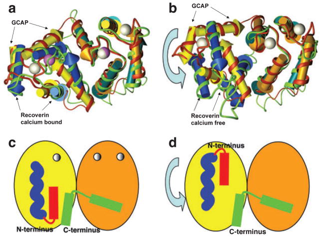
Photon absorption by rhodopsin triggers the phototransduction signaling pathway that culminates in degradation of cGMP, closure of cGMP-gated ion channels and hyperpolarization of the photoreceptor membrane. This process is accompanied by a decrease in free Ca(2+) concentration in the photoreceptor cytosol sensed by Ca(2+)-binding proteins that modulate phototransduction and activate the recovery phase to reestablish the photoreceptor dark potential. Guanylate cyclase-activating proteins (GCAPs) belong to the neuronal calcium sensor (NCS) family and are responsible for activating retinal guanylate cyclases (retGCs) at low Ca(2+) concentrations triggering synthesis of cGMP and recovery of the dark potential. Here we review recent structural insight into the role of the N-terminal myristoylation in GCAPs and compare it to other NCS family members. We discuss previous studies identifying regions of GCAPs important for retGC1 regulation in the context of the new structural data available for myristoylated GCAP1. In addition, we present a hypothetical model for the Ca(2+)-triggered conformational change in GCAPs and retGC1 regulation. Finally, we briefly discuss the involvement of mutant GCAP1 proteins in the etiology of retinal degeneration as well as the importance of other Ca(2+) sensors in the modulation of phototransduction.


Figures

Figure 1
Figure 1
Structure of myristoylated chicken GCAP1 with Ca2+ bound. The cartoon representation is colored yellow for EF-hands 1 and 2 and orange for EF-hands 3 and 4. The N-terminal helix is colored red, the kinked C-terminal helix is green and the myristoyl group is shown as a space-filling model in blue. Bound Ca2+ ions are shown in dark green.
Figure 2
Figure 2
Superposition of representative NCS protein structures in the Ca2+-bound form. Front (a) and back (b) cartoon representations of myrGCAP1 superimposed onto the NCS family members myr-recoverin, neurocalcin, NCS-1 and KChIP1. The coloring of myrGCAP1 is as in Fig. 1 with the N- and C-terminal helices in bright red and green, respectively. The other NCS proteins are colored light yellow for EF-hands 1 and 2 and light orange for EF-hands 3 and 4, dark red for the N-terminal helix and dark green for the C-terminal segment. The recoverin myristoyl group is shown as a space-filling model in light blue. Ca2+ ions bound to GCAP1 are shown in dark green for reference but are omitted for the other NCS proteins for clarity.
Figure 3
Figure 3
Surface residue conservation in GCAPs. Surface representations of myrGCAP1 in front (a), back (b), top (c) and bottom (d) orientations (a cartoon model oriented as in [a] is shown for reference). Residues conserved in GCAP1 across species, but not GCAP2, are colored blue. Residues conserved in GCAP1 and GCAP2, but not across the NCS family, are in red. Residues required for myristoylation and Ca2+ coordination across NCS family are shown in pink.
Figure 4
Figure 4
Regions of GCAP1 (a) and GCAP2 (b) identified in chimera studies as important for retGC regulation. Regions affecting retGC regulation are shown in red. The region of GCAP2 that reverses Ca2+ dependency is shown in cyan. The myristoyl group in GCAP1 is shown as a space-filling model in blue.
Figure 5
Figure 5
Model of the Ca2+-induced conformational change in GCAP1. (a) EF-hands of Ca2+-bound GCAP represented by yellow and orange arrowed cylinders, red ribbon and white calcium ions, superimposed on Ca2+-bound recoverin represented by blue and green-blue arrowed cylinders, green ribbon and violet Ca2+ ions. N- and C-terminal parts of the structure were removed for clarity. Superimposition was done on C-terminal EF-hand pairs (orange and green-blue cylinders). (b) EF-hands of Ca2+-bound GCAP superimposed on Ca2+-free recoverin. Superimposition was done on C-terminal EF-hand pairs. Coloring as in (a). (c, d) Schematic representation of a possible Ca2+-induced conformational change in GCAP1 where the N-terminal (red) and C-terminal (green) helices are clustered together in the Ca2+-bound state (c), but separated by domain rotation in the Ca2+-free form (d). The myristoyl group is represented in blue.

References

    1. Belgum JH, Copenhagen DR. Synaptic transfer of rod signals to horizontal and bipolar cells in the retina of the toad (Bufo marinus) J Physiol. 1988;396:225–245. - PMC - PubMed
    1. Palczewski K. G protein-coupled receptor rhodopsin. Annu Rev Biochem. 2006;75:743–767. - PMC - PubMed
    1. Palczewski K, Polans AS, Baehr W, Ames JB. Ca2+-binding proteins in the retina: Structure, function, and the etiology of human visual diseases. Bioessays. 2000;22:337–350. - PubMed
    1. Polans A, Baehr W, Palczewski K. Turned on by Ca2+! The physiology and pathology of Ca2+-binding proteins in the retina. Trends Neurosci. 1996;19:547–554. - PubMed
    1. Baehr W, Karan S, Maeda T, Luo DG, Li S, Bronson JD, Watt CB, Yau KW, Frederick JM, Palczewski K. The function of guanylate cyclase 1 and guanylate cyclase 2 in rod and cone photoreceptors. J Biol Chem. 2007;282:8837–8847. - PMC - PubMed

Publication types

LinkOut - more resources