. 2008 Mar 17:6:13.
doi: 10.1186/1479-5876-6-13.

Hypermethylated MAL gene - a silent marker of early colon tumorigenesis

Affiliations

Hypermethylated MAL gene - a silent marker of early colon tumorigenesis

Guro E Lind et al. J Transl Med. .

Abstract

Background: Tumor-derived aberrantly methylated DNA might serve as diagnostic biomarkers for cancer, but so far, few such markers have been identified. The aim of the present study was to investigate the potential of the MAL (T-cell differentiation protein) gene as an early epigenetic diagnostic marker for colorectal tumors.

Methods: Using methylation-specific polymerase chain reaction (MSP) the promoter methylation status of MAL was analyzed in 218 samples, including normal mucosa (n = 44), colorectal adenomas (n = 63), carcinomas (n = 65), and various cancer cell lines (n = 46). Direct bisulphite sequencing was performed to confirm the MSP results. MAL gene expression was investigated with real time quantitative analyses before and after epigenetic drug treatment. Immunohistochemical analysis of MAL was done using normal colon mucosa samples (n = 5) and a tissue microarray with 292 colorectal tumors.

Results: Bisulphite sequencing revealed that the methylation was unequally distributed within the MAL promoter and by MSP analysis a region close to the transcription start point was shown to be hypermethylated in the majority of colorectal carcinomas (49/61, 80%) as well as in adenomas (45/63, 71%). In contrast, only a minority of the normal mucosa samples displayed hypermethylation (1/23, 4%). The hypermethylation of MAL was significantly associated with reduced or lost gene expression in in vitro models. Furthermore, removal of the methylation re-induced gene expression in colon cancer cell lines. Finally, MAL protein was expressed in epithelial cells of normal colon mucosa, but not in the malignant cells of the same type.

Conclusion: Promoter hypermethylation of MAL was present in the vast majority of benign and malignant colorectal tumors, and only rarely in normal mucosa, which makes it suitable as a diagnostic marker for early colorectal tumorigenesis.


Figures

Figure 1
Figure 1
Methylation status of the MAL promoter in normal colon mucosa samples and colorectal carcinomas. Representative results from methylation-specific polymerase chain reaction are shown. A visible PCR product in lanes U indicates the presence of unmethylated alleles whereas a PCR product in lanes M indicates the presence of methylated alleles. N, normal mucosa; C, carcinoma; Pos, positive control (unmethylated reaction: DNA from normal blood, methylated reaction: in vitro methylated DNA); Neg, negative control (containing water as template); U, lane for unmethylated MSP product; M, lane for methylated MSP product.
Figure 2
Figure 2
Site specific methylation within the MAL promoter. Bisulphite sequencing of the MAL promoter verifies methylation status assessed by methylation-specific polymerase chain reaction. The upper part of the figure is a schematic presentation of the CpG sites successfully amplified by the two analyzed bisulphite sequencing fragments, A (-68 to +168; to the right) and B (-427 to -85; to the left). The transcription start site is represented by +1 and the vertical bars indicate the location of individual CpG sites. The two arrows indicate the location of the MSP primers in the present study and a previously published study analyzing promoter methylation of MAL [31]. For the lower part of the figure, filled circles represent methylated CpGs; open circles represent unmethylated CpGs; and open circles with a slash represent partially methylated sites (the presence of approximately 20–80% cytosine, in addition to thymine). The column of U, M and U/M at the right side of this lower part lists the methylation status of the respective cell lines as assessed by us using MSP analyses. Abbreviations: MSP, methylation-specific PCR; s, sense; as, antisense; U, unmethylated; M, methylated; U/M, presence of both unmethylated and methylated band.
Figure 3
Figure 3
The "bisulphite sequence" of the MAL promoter. Representative bisulphite sequencing electropherograms of the MAL promoter in colon cancer cell lines. A subsection of the bisulphite sequence electropherogram, covering CpG sites +11 to +15 relative to transcription start. Cytosines in CpG sites are indicated by a black arrow, whereas cytosines that have been converted to thymines are underlined in red. The MAL promoter sequencing electropherograms illustrated here, are from the unmethylated V9P cell line and the hypermethylated ALA and HCT116.
Figure 4
Figure 4
MAL expression in cancer cell lines and colorectal carcinomas. Promoter hypermethylation of MAL was associated with reduced or lost gene expression in in vitro models. The quantitative gene expression level of MAL is displayed as a ratio between the average of two MAL assays (detecting various splice variants) and the average of the two endogenous controls, GUSB and ACTB. The value has been multiplied by a factor of 1000. Below each sample the respective methylation status is shown, as assessed by methylation-specific polymerase chain reaction. Filled circles represent promoter hypermethylation of MAL, open circles represent unmethylated MAL, and open circles with a slash represent the presence of both unmethylated and methylated alleles. Colorectal carcinomas are divided in an unmethylated group (n = 3) and a hypermethylated group (n = 13), and the median expression is displayed here. The tissue of origin for the individual cell lines can be found in table 1.
Figure 5
Figure 5
Up-regulation of MAL expression after drug treatment. Decreased promoter methylation of MAL followed by up-regulated mRNA expression in colon cancer cell lines was found after treatment with the demethylating 5-aza-2'deoxycytidine, alone and in combination with the deacetylase inhibitor trichostatin A. Upper panel demonstrate the relative expression values of MAL (linear scale) in two colon cancer cell lines, HT29 and HCT15, treated with 5-aza-2'deoxycytidine alone, trichostatin A alone, and the two drugs in combination. Lower panel illustrate MAL MSP results for the same samples. A visible PCR product in lanes U indicates the presence of unmethylated alleles whereas a PCR product in lanes M indicates the presence of methylated alleles. Abbreviation: AZA, 5-aza-2'deoxycytidine; TSA, trichostatin A; Pos, positive control (unmethylated reaction: DNA from normal blood, methylated reaction: in vitro methylated DNA); Neg, negative control (containing water as template); U, lane for unmethylated MSP product; M, lane for methylated MSP product.
Figure 6
Figure 6
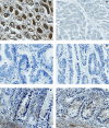
Lack of MAL protein expression in colorectal carcinomas. Positive cytoplasmic staining of MAL was found in kidney tubuli (A), and no staining was observed in heart muscle (B), in agreement with earlier reports [30]. The epithelial cells of colorectal carcinomas were MAL negative (C, D), whereas in normal colon tissue, cytoplasmic expression of MAL was found in both epithelia and connective tissue (E, F). All images were captured using the 40× lens (400× magnification).

References

    1. Feinberg AP, Ohlsson R, Henikoff S. The epigenetic progenitor origin of human cancer. Nat Rev Genet. 2006;7:21–33. doi: 10.1038/nrg1748. - DOI - PubMed
    1. Kane MF, Loda M, Gaida GM, Lipman J, Mishra R, Goldman H, Jessup JM, Kolodner R. Methylation of the hMLH1 promoter correlates with lack of expression of hMLH1 in sporadic colon tumors and mismatch repair-defective human tumor cell lines. Cancer Res. 1997;57:808–811. - PubMed
    1. Herman JG, Umar A, Polyak K, Graff JR, Ahuja N, Issa JP, Markowitz S, Willson JK, Hamilton SR, Kinzler KW, Kane MF, Kolodner RD, Vogelstein B, Kunkel TA, Baylin SB. Incidence and functional consequences of hMLH1 promoter hypermethylation in colorectal carcinoma. Proc Natl Acad Sci U S A. 1998;95:6870–6875. doi: 10.1073/pnas.95.12.6870. - DOI - PMC - PubMed
    1. Cunningham JM, Christensen ER, Tester DJ, Kim CY, Roche PC, Burgart LJ, Thibodeau SN. Hypermethylation of the hMLH1 promoter in colon cancer with microsatellite instability. Cancer Res. 1998;58:3455–3460. - PubMed
    1. Esteller M, Sparks A, Toyota M, Sanchez-Cespedes M, Capella G, Peinado MA, Gonzalez S, Tarafa G, Sidransky D, Meltzer SJ, Baylin SB, Herman JG. Analysis of adenomatous polyposis coli promoter hypermethylation in human cancer. Cancer Res. 2000;60:4366–4371. - PubMed

Publication types

MeSH terms