. 2008 Jun;1783(6):1261-70.
doi: 10.1016/j.bbamcr.2008.02.004. Epub 2008 Feb 20.

Role of the carboxyl terminal di-leucine in phosphorylation and internalization of C5a receptor

Affiliations

Role of the carboxyl terminal di-leucine in phosphorylation and internalization of C5a receptor

Elena S Suvorova et al. Biochim Biophys Acta. 2008 Jun.

Abstract

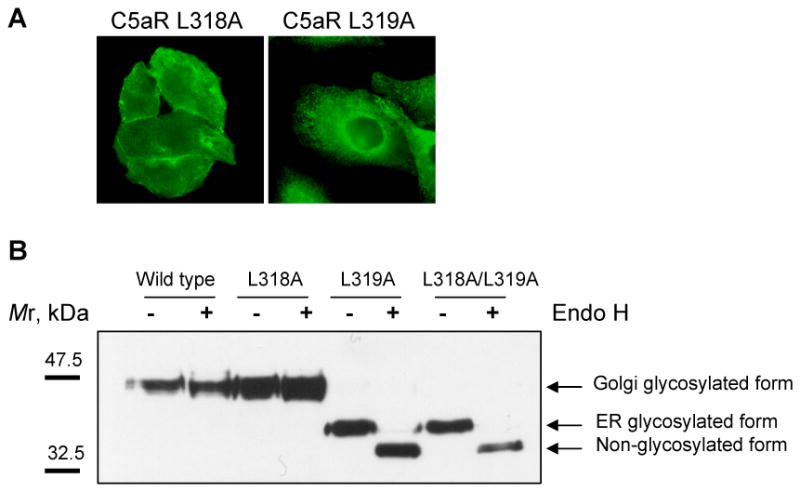
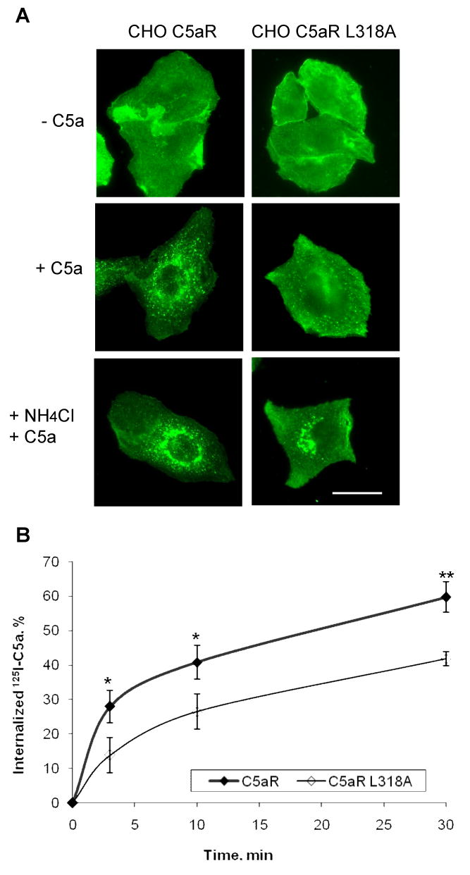
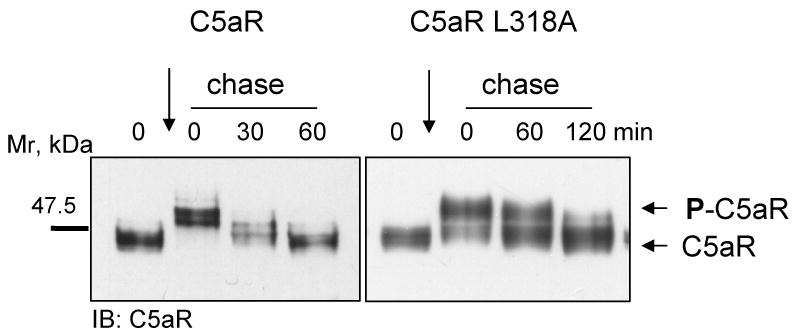
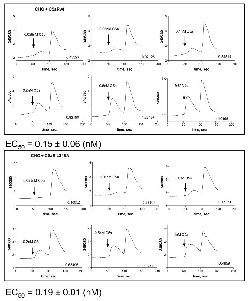
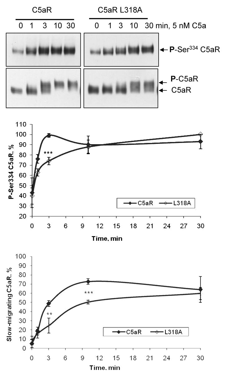
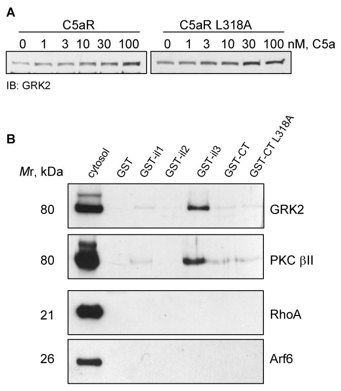
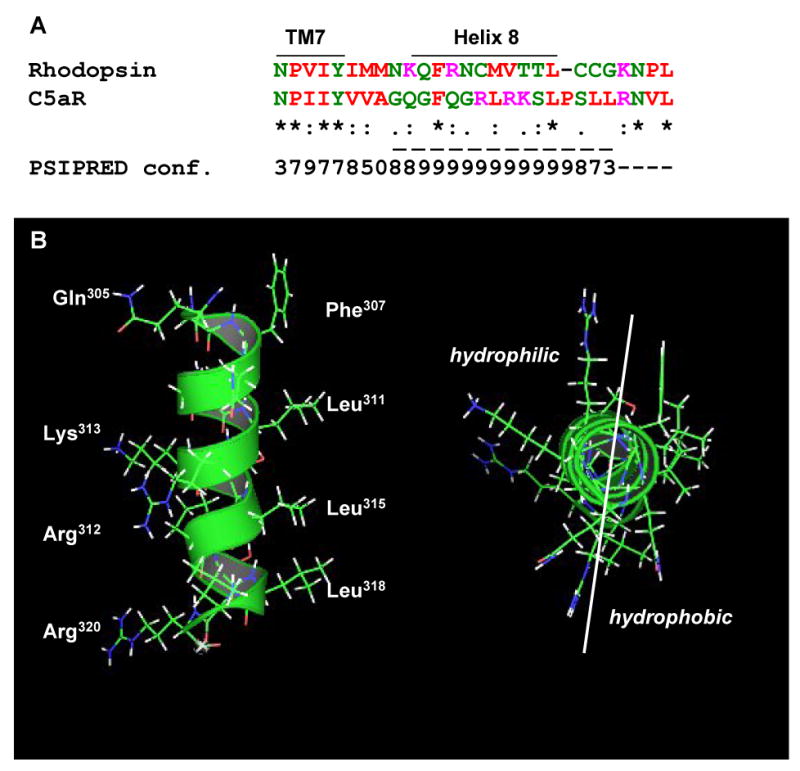
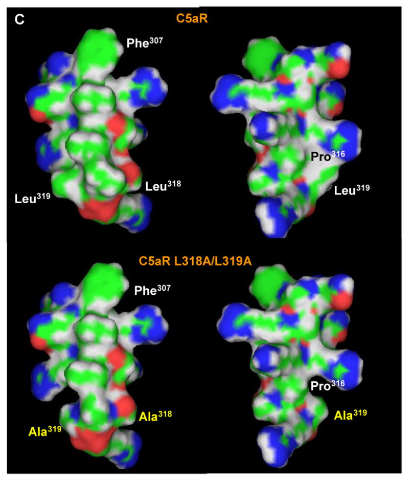
The carboxyl tail of G protein-coupled receptors contains motifs that regulate receptor interactions with intracellular partners. Activation of the human neutrophil complement fragment C5a receptor (C5aR) is terminated by phosphorylation of the carboxyl tail followed by receptor internalization. In this study, we demonstrated that bulky hydrophobic residues in the membrane-proximal region of the C5aR carboxyl tail play an important role in proper structure and function of the receptor: Substitution of leucine 319 with alanine (L319A) resulted in receptor retention in the endoplasmic reticulum, whereas a L318A substitution allowed receptor transport to the cell surface, but showed slow internalization upon activation, presumably due to a defect in phosphorylation by both PKC and GRK. Normal agonist-induced activation of ERK1/2 and intracellular calcium release suggested that the L318A mutation did not affect receptor signaling. Binding of GRK2 and PKCbetaII to intracellular loop 3 of C5aR in vitro indicated that mutagenesis of L318 did not affect kinase binding. Limited proteolysis with trypsin revealed a conformational difference between wild type and mutant receptor. Our studies support a model in which the L318/L319 stabilizes an amphipathic helix (Q305-R320) in the membrane-proximal region of C5aR.


Figures

Figure 1
Figure 1. Exchange of L319 to alanine resulted in retention of C5aR in the ER
A) Immunofluorescence microscopy of stably transfected CHO cells expressing either C5aR L318A or C5aR L319A. Immunostaining with anti-C5aR antibody showed plasma membrane localization of C5aR L318A and intracellular retention of C5aR L319A. The images are representative of four different clones for each mutant receptor. B) Analysis of susceptibility of wild type C5aR and di-leucine mutants to Endo H treatment. Membranes of CHO cells expressing either wild type or mutant C5aR were denatured for 10 min at 60°C and treated with Endo H according to the manufacturer's recommendations (New England Biolabs Inc.). Non-treated (-) and Endo H treated (+) membranes were separated in 10% SDS-polyacrylamide gel and analyzed by western blotting using anti-C5aR antibody. Arrows indicate Golgi-glycosylated, ER-glycosylated and non-glycosylated forms of C5aR. One representative image is shown.
Figure 2
Figure 2. Internalization of C5aR L318A occurs at a reduced rate
A) Stable CHO transfectants were incubated for 3 h with or without 100 nM C5a. To accumulate internalized receptor, cells were incubated with ligand and 50 mM NH4Cl. Receptors were visualized using rabbit anti-C5aR antibody. Representative images of two independent experiments are shown. Bar 50 μm. B) CHO transfectants were incubated at 37°C with 125I-C5a for the indicated times. Surface bound ligand was removed by washing cells with cold PBS, pH 2.8. Internalization was calculated as the percentage of the acid resistant radioactivity out of total cell-associated radioactivity. Results are mean ± S.D. of three independent experiments. (*, p < 0.05; **, p < 0.01).
Figure 3
Figure 3. C5aR L318A becomes dephosporylated at a slower rate than wild-type C5aR
Cells were incubated for 10 min at 37°C with 50 nM C5a followed by a 0, 30 or 60 min chase in warm medium containing 1 mM cycloheximide (to inhibit protein synthesis). Wild type C5aR reached complete dephosphorylation after 60 min, whereas C5aR L318A remained partly phosphorylated up to 120 min.
Figure 4
Figure 4. Wild type and mutant C5aR L318A showed similar activation of ERK1/2
A) CHO transfectants were pre-incubated overnight with or without 200 ng/ml PTX and activated with 100 nM C5a for the indicated times. ERK1/2 phosphorylation was detected in the cell lysates by immunoblotting using anti-phospho-ERK1/2 antibody. Samples were tested for equal loading and expression of ERK1/2 by immunoblotting with anti-ERK1/2 antibody. B) CHO transfectants were exposed for 5 min to the indicated concentrations of C5a. Anti-phospho-ERK1/2 antibody was used to detect phosphorylated ERK1/2. Equal loading was tested by staining the filters with Ponceau S prior to antibody incubation (data not shown).
Figure 5
Figure 5. Wild type and C5aR L318A mutant induced similar C5a-mediated mobilization of intracellular Ca2+
CHO transfectants were loaded with Fura 2-AM and calcium release was measured in dual wavelength excitation in response to addition of various concentrations of C5a (added at 50 sec). 10 μM ATP was added at 100 sec as a positive control stimulus. Numbers on each graph shows the difference between the base line value and the C5a-induced peak value. EC50 was calculated from three independent experiments by non-linear regression analysis.
Figure 6
Figure 6. Mutation of L318 led to attenuated phosphorylation of C5aR
CHO cells stably expressing C5aR or C5aR L318A were stimulated with 5 nM C5a for the indicated times. Dynamics of the S334 phosphorylation were analyzed by western blotting of the total membranes using anti-phosphoS334-C5aR monoclonal antibody 32-G1 (upper panel). GRK-mediated phosphorylation was analyzed from the same samples by western blotting using polyclonal C5aR antibody (lower panel). The graphs show the relative amounts of phosphorylated C5aR. In the upper graph, the data are expressed as the percentage of S334 phosphorylation with the strongest signal set at 100%. In the lower graph, the relative amount of phosphorylated C5aR is shown as the percentage of slow-migrating C5aR out of total C5aR. Since these density measurements are difficult to carry out with complete accuracy, the results should not be viewed as absolute, but indicative of the phosphorylation trend. Each data point represents the mean value ± S.D. of three independent experiments. (**, p < 0.01; ***, p < 0.001)
Figure 7
Figure 7
A) GRK2 translocates to the cell membrane upon activation with C5a. CHO transfectants were stimulated for 10 min with the indicated amounts of C5a, and GRK2 was detected from membrane fractions by western blotting. One representative image from three experiments is shown. B) PKCβII and GRK2 bind preferentially to the third intracellular loop of C5aR in in vitro binding assay. Affinity purified GST-tagged cytoplasmic domains of C5aR and the carboxyl tail of C5aR L318A were incubated with cytosol from activated neutrophils. Eluates were analyzed by immunoblotting with anti-GRK2, anti-phospho-PKCβII, anti-RhoA, and anti-Arf6 antibody. 2% of the cytosol versus 25% of the eluate was loaded. Abbreviation: il - intracellular loop, CT – carboxyl tail.
Figure 8
Figure 8. Limited proteolysis of C5aR and C5aR L318A in CHO membranes
Triton X-100 solubilized membranes of CHO cells expressing C5aR or C5aR L318A were treated with trypsin. Samples were analyzed in western blots using anti-C5aR antibody. Images are representative of three independent experiments. The graph shows relative receptor degradation during 1 h incubation. Dynamics of the receptor disappearance on the western blots was analyzed by scanning densitometry using Scion Image software. Signal from non-treated membranes (0 min) was set at 100%. Each data point is the mean ± S.D. of three independent experiments.
Figure 9
Figure 9. A structural model of the membrane-proximal domain of C5aR
A) Sequence alignment of the putative helix 8 of bovine rhodopsin and human C5aR. PSIPRED prediction numbers represent the confidence rate for a helical secondary structure (0 = low, 9 = high). Dashed line underlines a cytoplasmic helix predicted by PSIPRED (http://bioinf.cs.ucl.ac.uk/psipred/). Color code: red - small and hydrophobic residues, magenta - basic residues, blue - acidic, green - hydroxyl, amine, basic residues and glutamine. B) Side view (left) and projection across the extended amphipathic helix 8 (right) of C5aR. C) Mutation of di-leucine L318/L319 to alanines significantly reduces the hydrophobicity around the COOH-terminus of the helix. Left and right panels show the hydrophobic and hydrophilic surfaces, respectively, of the C5aR (upper panel) and C5aR L318A/L319A (lower panel) helix. Leucine to alanine mutation is shown in yellow.

Similar articles

Cited by

References

    1. Rabiet MJ, Huet E, Boulay F. The N-formyl peptide receptors and the anaphylatoxin C5a receptors: an overview. Biochimie. 2007;89:1089. - PMC - PubMed
    1. Reiter E, Lefkowitz RJ. GRKs and β-arrestins: roles in receptor silencing, trafficking and signaling. Trends Endocrin Metab. 2006;17:159. - PubMed
    1. Loudon RP, Perussia B, Benovic JL. Differentially regulated expression of the G-protein-coupled receptor kinases, betaARK and GRK6, during myelomonocytic cell development in vitro. Blood. 1996;88:4547. - PubMed
    1. Langkabel P, Zwirner J, Oppermann M. Ligand-induced phosphorylation of anaphylatoxin receptors C3aR and C5aR is mediated by G protein-coupled receptor kinases. Eur J Immunol. 1999;29:3035. - PubMed
    1. Chuang TT, Sallese M, Ambrosini G, Parruti G, De Blasi A. High expression of β-adrenergic receptor kinase in human peripheral blood leukocytes. Isoproterenol and platelet activating factor can induce kinase translocation. J Biol Chem. 1992;267:6886. - PubMed

Publication types

MeSH terms

LinkOut - more resources