Comparative Study
. 2008 May;36(5):655-66.
doi: 10.1016/j.exphem.2008.01.002. Epub 2008 Mar 17.

Deficiency of Src family kinases compromises the repopulating ability of hematopoietic stem cells

Affiliations
Comparative Study

Deficiency of Src family kinases compromises the repopulating ability of hematopoietic stem cells

Christie M Orschell et al. Exp Hematol. 2008 May.

Abstract

Objective: Src family kinases (SFK) have been implicated in regulating growth factor and integrin-induced proliferation, migration, and gene expression in multiple cell types. However, little is known about the role of these kinases in the growth, homing, and engraftment potential of hematopoietic stem and progenitor cells.

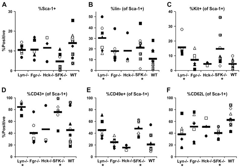
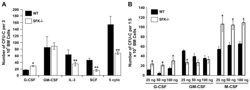
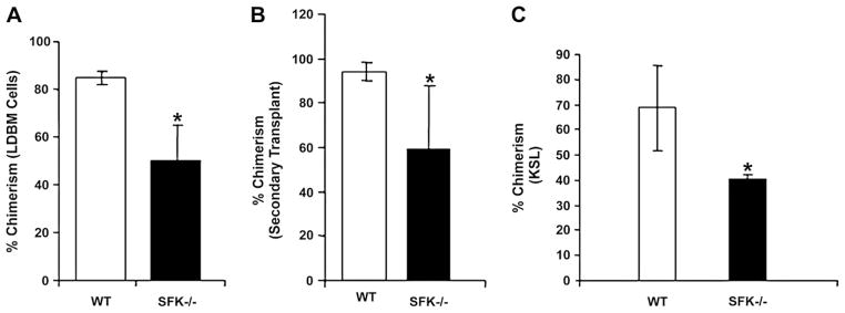
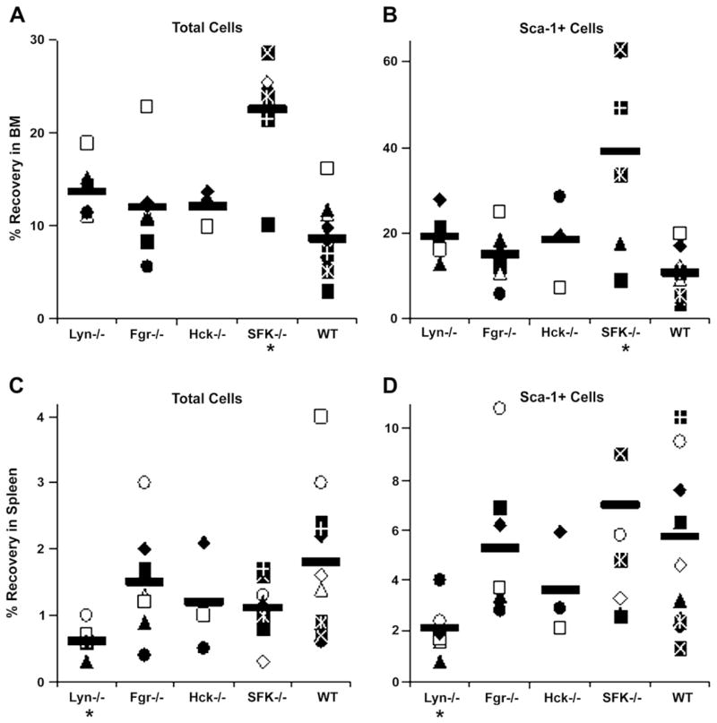
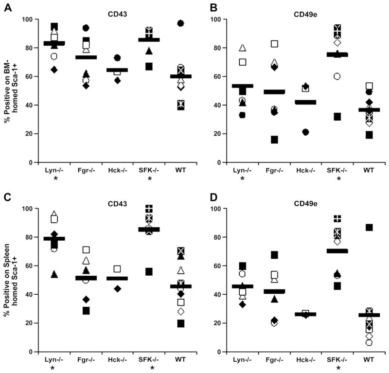
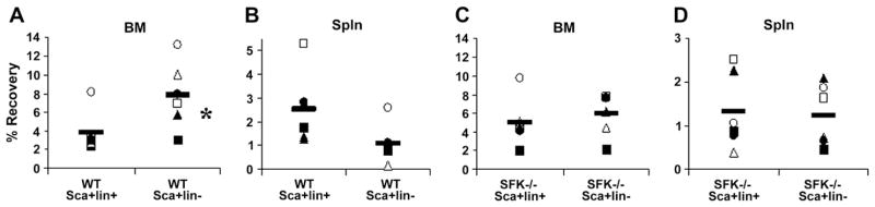
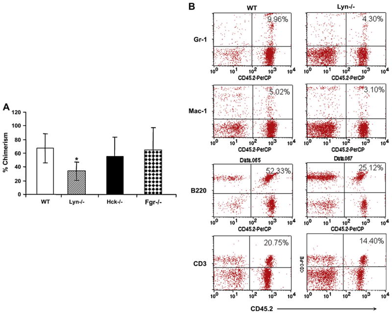
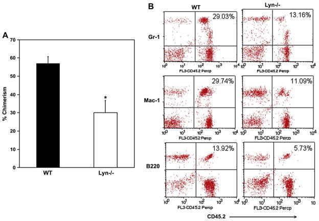
Results: Here we show that loss of hematopoietic-specific SFKs Hck, Fgr, and Lyn results in increased number of Sca-1(+)Lin(-) cells in the bone marrow, which respond differentially to cytokine-induced growth in vitro and manifest a significant defect in the long-term repopulating potential in vivo. Interestingly, a significant increase in expression of adhesion molecules, known to coincide with the homing potential of wild-type bone marrow cells is also observed on the surface of SFK(-/-) cells, although, this increase did not affect the homing potential of more primitive Lin(-)Sca-1(+) SFK(-/-) cells. The stem cell-repopulating defect observed in mice transplanted with SFK(-/-) bone marrow cells is due to the loss of Lyn Src kinase, because deficiency of Lyn, but not Hck or Fgr, recapitulated the long-term stem cell defect observed in mice transplanted with SFK(-/-) bone marrow cells.

Conclusions: Taken together, our results demonstrate an essential role for Lyn kinase in positively regulating the long-term and multilineage engraftment of stem cells, which is distinct from its role in mature B cells and myeloid cells.


Figures

Figure 1
Figure 1
Primitive phenotype of Src family kinase (SFK) knockout cells. Low-density bone marrow (LDBM) cells from SFK knockout mice were analyzed for percentage of Sca-1+ cells and markers known to be expressed on primitive hematopoietic cells. (A) Percentage of Sca-1+ cells within LDBM cells. Percentages of Sca-1+ cells lacking lineage markers (CD3, B220, Gr-1, B), or expressing KIT (C), CD43 (D), CD49e (E), or CD62L (F) are shown in the remaining graphs. Data are from individual mice in 3 to 11 separate experiments; mean is given by horizontal line. *p < 0.05 compared with wild-type (WT).
Figure 2
Figure 2
Deficiency of Src family kinases (SFKs) in bone marrow (BM) cells results in different responses to cytokines. Low-density BM (LDBM) cells from wild-type (WT) or SFK−/− mice were harvested and subjected to methylcellulose assay. (A) 3 × 10⁴ LDBM cells were grown in methylcellulose in the presence of granulocyte colony-stimulated factor (G-CSF; 25 ng/mL), granulocyte macrophage colony-stimulating factor (GM-CSF; 50 ng/mL), interleukin (IL)-3 (10 ng/mL), stem cell factor (SCF; 50 ng/mL), or a combination of five cytokines (erythropoietin 2 U/mL, G-CSF, SCF, IL-3, and GM-CSF). Colony-forming units in culture (CFU-Cs) were scored 7 to 10 days later. Bars represent the mean number of CFU-Cs ± standard deviation (SD) at various doses and in the presence of different cytokines. *p < 0.05 compared with WT; **p < 0.05 compared with WT. (B) 1.5 × 10⁴ LDBM cells were grown in methyl-cellulose in the presence of indicated concentrations of G-CSF, GM-CSF, and M-CSF. CFU-Cs were scored 7 to 10 days later. Bars represent the mean number of CFU-Cs ± SD at the indicated doses in the presence of different cytokines. *p < 0.05 compared to WT.
Figure 3
Figure 3
Deficiency of Src family kinase (SFK) results in impaired repopulating capacity in a competitive repopulation assay. Peripheral blood chimerism in mice transplanted with wild-type (WT) or SFK−/− low-density bone marrow (LDBM) (A) or KIT+LinSca-1+ cells (C) and 1 × 10⁵ competitor cells. Mean chimerism for LDBM (A) and c-Kit+LinSca-1+ (C) cell number transplanted was calculated for each genotype, and the data generated at 6 months after transplantation are summarized in bar graphs. Eight mice (four per genotype) were transplanted at each test cell concentration (three different concentrations were used: 1:1, 1:2, and 1:3 for LDBM cells and two different concentrations were used for Kit+LinSca-1+ cells (1000 and 10,000). Experiments using LDBM were repeated twice. A total of 32 mice were transplanted (16 per genotype). Shown are data from 1:1 ratio in LDBM and 10,000 KIT+Lin–Sca-1+ cells. Mean ± standard deviation (SD), *p < 0.05 SFK−/− vs WT. (B) LDBM cells from lethally irradiated primary recipients transplanted with WT or SFK−/− BM that demonstrated the highest chimerism (1:3; competitor-to-test ratio) were harvested and 1 × 10⁶ cells were transplanted into lethally irradiated secondary hosts. Mean chimerism was calculated for each genotype and data generated at 6 months after secondary transplantation are summarized in bar graphs. BM from 2 independent WT and SFK−/− primary recipients were transplanted into 24 lethally irradiated (12 for each genotype) mice. Mean ± SD, *p < 0.05, SFK−/− vs WT. KSL = KIT+Sca-1+Lin.
Figure 4
Figure 4
Percent recovery of transplanted Src family kinases (SFK) knockout cells in bone marrow (BM) and spleen. Irradiated wild-type (WT) mice received 17.5 × 10⁶ to 20 × 106 low-density BM cells isolated from Lyn−/−, Fgr−/−, Hck−/−, SFK−/−, or WT mice, sacrificed 16 to 19 hours later, and percent recovery of total donor cells and donor Sca-1+ cells in harvested BM (A, B) and spleen (C, D) were calculated as described in Materials and Methods. Data represent percent recovery in individual mice in 3 to 11 separate experiments; mean is given by horizontal line. *p < 0.05 compared with WT.
Figure 5
Figure 5
Frequency of CD43+ and CD49e+ cells among donor Sca-1+ cells homed to bone marrow (BM) or spleen after transplantation. Irradiated wild-type (WT) mice received 17.5 × 10⁶ to 20 × 106 low-density LDBM cells isolated from Lyn−/−, Fgr−/−, Hck−/−, Src family kinases (SFK)−/−, or wild-type (WT) mice, sacrificed 16 to 19 hours later, and expression of CD43 and CD49e examined on donor Sca-1+ cells recovered in BM (A, B) and spleen (C, D). Data represent percentage of recovered donor Sca-1+ cells expressing CD43 or CD49e in individual mice in four to nine separate experiments; mean is given by horizontal line. *p < 0.05 compared with WT.
Figure 6
Figure 6
Percent recovery of transplanted Src family kinase (SFK)−/− or wild-type (WT) Sca-1+lin+ or Sca-1+lin graft cells in bone marrow (BM) and spleen of WT recipients. Irradiated WT mice received 1.4 × 10⁵ to 1.6 0 105 purified Sca-1+lin+ or Sca-1+lin cells isolated from SFK−/− or WT mice, sacrificed 16 to 19 hours later, and percent recovery of total donor cells in harvested BM (A, C) and spleen (B, D) was calculated as described in Materials and Methods. Data represent % recovery in individual mice in four separate experiments (n = 6 mice total); horizontal line represents mean. *p < 0.05 compared with WT.
Figure 7
Figure 7
Competitive repopulating ability of hematopoietic stem/progenitor (HSC/P) cells lacking expression of Hck, Fgr, or Lyn. (A) Peripheral blood (PB) lineage chimerism in mice transplanted with wild-type (WT), Hck−/−, Lyn−/−, or Fgr−/− low-density bone marrow (BM) cells and 1 × 10⁵ competitor cells. Mean chimerism for each genotype was calculated and the data generated at 4 months after transplantation are summarized in bar graphs. Experiments were repeated twice with four to five mice in each group. Shown are data from 1:1 ratio. Mean ± standard deviation, *p < 0.05 WT vs Lyn−/−. (B) Representative dot blots indicating multilineage engraftment of WT and Lyn−/− stem cells into myeloid and lymphoid lineages. PB cells were stained with antibodies that recognize myeloid and lymphoid lineages. Shown are percentages of donor cells from a representative mouse engrafted with WT or Lyn−/− BM cells and competitors.
Figure 8
Figure 8
Reduced engraftment of Lyn−/− stem cells in lethally irradiated secondary recipients. Bone marrow (BM) of wild-type (WT) or Lyn−/− primary recipients were harvested after 4 months of primary transplantation and 1 × 10⁶ low-density BM (LDBM) cells were transplanted into lethally irradiated secondary hosts. (A) Shows the mean chimerism for each genotype after 12 weeks post secondary transplantation. LDBM cells from a WT and Lyn−/− primary recipient were transplanted into four to five lethally irradiated secondary recipients. Mean ± SD, *p < 0.05, Lyn−/− vs WT. (B) Representative dot blots indicating myeloid and lymphoid lineage engraftment in secondary recipients. Shown are percentages of donor cells from a representative mouse engrafted with WT or Lyn−/− LDBM cells.

References

    1. Lannutti BJ, Drachman JG. Lyn tyrosine kinase regulates thrombopoietin-induced proliferation of hematopoietic cell lines and primary megakaryocytic progenitors. Blood. 2004;103:3736–3743. - PubMed
    1. Lannutti BJ, Minear J, Blake N, Drachman JG. Increased megakaryo-cytopoiesis in Lyn-deficient mice. Oncogene. 2006;25:3316–3324. - PubMed
    1. Lannutti BJ, Shim MH, Blake N, Reems JA, Drachman JG. Identification and activation of Src family kinases in primary megakaryocytes. Exp Hematol. 2003;31:1268–1274. - PubMed
    1. Borneo J, Munugalavadla V, Sims EC, et al. Src family kinase-mediated negative regulation of hematopoietic stem cell mobilization involves both intrinsic and microenvironmental factors. Exp Hematol. 2007;35:1026–1037. - PMC - PubMed
    1. Mermel CH, McLemore ML, Liu F, et al. Src family kinases are important negative regulators of G-CSF-dependent granulopoiesis. Blood. 2006;108:2562–2568. - PMC - PubMed

Publication types