. 2008 May 16;283(20):13638-51.
doi: 10.1074/jbc.M802027200. Epub 2008 Mar 17.

Roles of Pofut1 and O-fucose in mammalian Notch signaling

Affiliations

Roles of Pofut1 and O-fucose in mammalian Notch signaling

Mark Stahl et al. J Biol Chem. .

Abstract

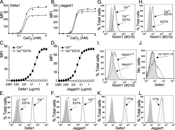
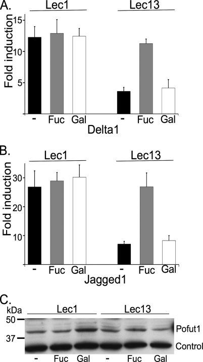
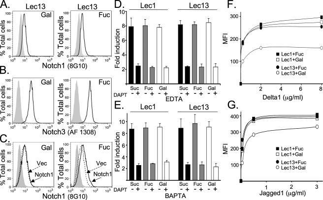
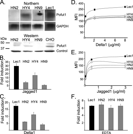
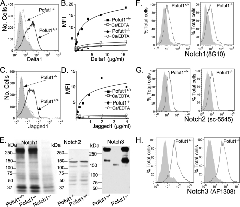
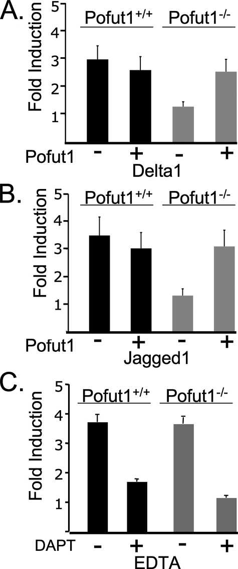
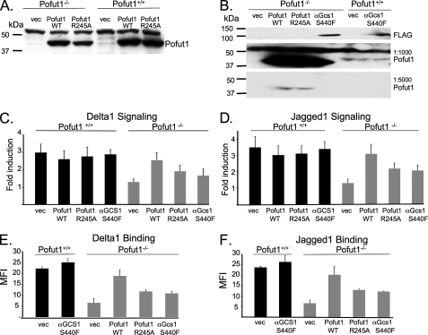
Mammalian Notch receptors contain 29-36 epidermal growth factor (EGF)-like repeats that may be modified by protein O-fucosyltransferase 1 (Pofut1), an essential component of the canonical Notch signaling pathway. The Drosophila orthologue Ofut1 is proposed to function as both a chaperone required for stable cell surface expression of Notch and a protein O-fucosyltransferase. Here we investigate these dual roles of Pofut1 in relation to endogenous Notch receptors of Chinese hamster ovary and murine embryonic stem (ES) cells. We show that fucosylation-deficient Lec13 Chinese hamster ovary cells have wild type levels of Pofut1 and cell surface Notch receptors. Nevertheless, they have reduced binding of Notch ligands and low levels of Delta1- and Jagged1-induced Notch signaling. Exogenous fucose but not galactose rescues both ligand binding and Notch signaling. Murine ES cells lacking Pofut1 also have wild type levels of cell surface Notch receptors. However, Pofut1-/- ES cells do not bind Notch ligands or exhibit Notch signaling. Although overexpression of fucosyltransferase-defective Pofut1 R245A in Pofut1-/- cells partially rescues ligand binding and Notch signaling, this effect is not specific. The same rescue is achieved by an unrelated, inactive, endoplasmic reticulum glucosidase. Therefore, mammalian Notch receptors require Pofut1 for the generation of optimally functional Notch receptors, but, in contrast to Drosophila, Pofut1 is not required for stable cell surface expression of Notch. Importantly, we also show that, under certain circumstances, mammalian Notch receptors are capable of signaling in the absence of Pofut1 and O-fucose.


Figures

FIGURE 1.
FIGURE 1.
Notch ligand binding to CHO cells. A, Notch ligand binding is dependent on Ca2+ concentration. Binding of 2 μg/ml (filled circles) and 0.5 μg/ml (open circles) soluble Delta1-Fc to Lec1 cells over a range of calcium concentrations was determined by flow cytometry. Data are MFI with ligand and secondary antibody minus MFI with secondary Ab alone. The optimal least squares fits of Hill's equation (Y = A × XN/(K0.5N + XN)) (solid lines) are shown. B, binding of 0.5 μg/ml (filled circles) and 0.125 μg/ml (open circles) soluble Jagged1-Fc to Lec1 cells as in A. C, binding of soluble Delta1-Fc to Lec1 cells over a range of Delta1-Fc concentrations by flow cytometry. Filled circles, incubation in buffer containing 1 mm CaCl2; open circles, incubation in buffer containing 1 mm CaCl2 and 5 mm EDTA. Data are MFI with ligand and secondary antibody minus MFI with secondary Ab alone. D, binding of Jagged1-Fc to Lec1 cells as in C. E, binding of 8 μg/ml soluble Delta1-Fc to Lec1 cells analyzed by flow cytometry. The shaded profile is secondary antibody alone, the solid profile is for ligand in buffer containing 1 mm CaCl2, and the dashed profile is ligand binding in buffer containing 1 mm CaCl2 and 5 mm EDTA. F, binding of 4 μg/ml soluble Jagged1-Fc to Lec1 cells as in E. G, anti-Notch1 ECD antibody 8G10 binding to Lec1 cells incubated in binding buffer with 1 mm CaCl2 (solid line) or 1 mm CaCl2 and 5 mm EDTA (dashed line). H, Notch1 antibody binding to Lec1 cells in buffer containing 5 mm EDTA with 1 mm CaCl2 (solid line) or EDTA without calcium (dashed line). The shaded profile is for secondary antibody alone. I, antibody 8G10 is specific for Notch1. Anti-Notch1 ECD antibody 8G10 binding to Notch1+/+ ES cells (solid line) compared with Notch1–/– ES cells (dashed line) was analyzed by flow cytometry. The shaded profile is secondary antibody alone. J, a Notch1 ECD fragment inhibits Delta1-Fc binding. Notch1 fragment EGF-(1–18) (N1–18) prepared in Lec1 CHO cells was preincubated with soluble Delta1-Fc (0.5 μg/ml) for 30 min before the mixture was incubated with Pofut1+/+ ES cells and analyzed by flow cytometry (solid line). The dashed profile is binding of Delta1-Fc in the absence of N1–18, and the shaded profile is for secondary antibody alone. K, ligand binding to Lec1 cells is modulated by Fringe. The shaded profile is for secondary antibody alone, the solid profile is for binding of 2 μg/ml soluble Delta1-Fc to Lec1 cells stably expressing Lfng, and the dashed line is for binding of Delta1-Fc to Lec1 cells stably expressing control vector. L, soluble Jagged1-Fc (0.5 μg/ml) binding to Lec1 cells expressing Lfng (solid line) or control vector (dashed line).
FIGURE 2.
FIGURE 2.
Lec13 cells are deficient in ligand-induced Notch signaling but express Pofut1. Lec1 and Lec13 cells were cultured in medium containing no addition (–) or 1 mm fucose (Fuc) or 1 mm galactose (Gal) for 4 days. A, Delta1-induced Notch signaling in Lec1 and Lec13 cells. -Fold induction for Delta1/L:L was calculated after normalization. Error bars, S.D. (n = 6). B, Jagged1-induced Notch signaling in Lec1 and Lec13 cells. -Fold induction for Jagged1/L:L was calculated after normalization. Error bars, S.D. (n = 6). C, cell lysates (50 μg of protein) were analyzed by Western blot using bovine anti-Pofut1 antibodies (diluted 1:500). Control is a nonspecific band on the same blot.
FIGURE 3.
FIGURE 3.
Fucose supplementation rescues Notch ligand binding but does not affect cell surface expression of endogenous Notch receptors in Lec13 cells. Lec13 cells grown in medium supplemented with 1 mm fucose or 1 mm galactose were incubated with Notch1 extracellular domain antibody (8G10) (A) or Notch3 extracellular domain antibody (AF1308) (B) and analyzed by flow cytometry. The shaded profiles are secondary antibody alone. C, Lec13 cells cultured in 1 m m Gal or 1 mm Fuc were transfected with Notch1 (solid profile) or empty vector (dashed profile) and analyzed for binding of anti-Notch1 ECD antibody 8G10 by flow cytometry. D, ligand-independent signaling following calcium depletion by EDTA. Cells were cultured for 4 days in medium containing 1 mm fucose, 1 mm galactose, or 1 mm sucrose and cotransfected with the TP-1 Notch-responsive luciferase and control pRL-TK Renilla luciferase reporters. After 16 h, cells were incubated in Hanks' balanced salt solution containing 4 mm EDTA or 2.5 mm CaCl2 with or without the γ-secretase inhibitor DAPT (500 nm) for 5 min and then in complete medium for 6 h before assessing Notch activation by a dual luciferase assay. -Fold induction is the ratio of normalized EDTA-treated to CaCl2-treated cells. Error bars, S.D. (n = 4). E, ligand-independent signaling following calcium depletion by 7.5 mm BAPTA performed as described in D. F, Lec13 cells are deficient in Notch ligand binding and rescued by fucose. Lec1 cells (squares) and Lec13 cells (circles) were cultured for 4 days in 1 mm fucose (solid) or 1 mm galactose (open) and tested for soluble Delta1-Fc binding by flow cytometry. Each data point indicates MFI of primary and secondary antibody minus binding to secondary Ab alone. G, soluble Jagged1 binding to Lec1 and Lec13 cells performed as described in F.
FIGURE 4.
FIGURE 4.
CHO cells deficient in Pofut1 have reduced Notch signaling and ligand binding. A, total RNA from Lec1 or Lec1 cell lines stably expressing RNAi targeting Pofut1 transcripts was analyzed by a Northern blot hybridized to a probe from the Pofut1 gene coding region (top). The same blot was stripped and hybridized to glyceraldehyde-3-phosphate dehydrogenase (GAPDH). The star indicates a nonspecific signal. Detergent lysates prepared from the same cell lines were analyzed by Western blot for Pofut1 protein (bottom). Control is a nonspecific band on the same gel. B, Lec1 cells expressing RNAi targeted against Pofut1 transcripts were assayed for Notch signaling induced by co-culture with Jagged1/L compared with control L cells. Bars, -fold induction of signaling with S.D. (n = 4). C, Lec1 cells expressing RNAi targeted against Pofut1 transcripts were assayed for Delta1-induced Notch signaling as in B. D, binding of Delta1-Fc to parental Lec1 (filled squares), HY4 (open circles), HN2 (open triangles), and HN9 (open diamonds) in RNAi-targeted cells analyzed by flow cytometry. MFI is given after subtraction of MFI for secondary antibody alone. E, binding of Jagged1-Fc to Lec1 (filled squares), HY4 (open circles), HN2 (open triangles), and HN9 (open diamonds) by flow cytometry analyzed as in D. F, ligand-independent signaling in Lec1 cells with RNAi-targeted Pofut1 and parental Lec1 cells following incubation in 4 mm EDTA or 2.5 mm CaCl2 for 5 min followed by culture for 8 h. -Fold induction is given as the ratio of normalized luciferase activity in EDTA-treated compared with CaCl2-treated cells. Bars, S.D. (n = 4).
FIGURE 5.
FIGURE 5.
ES cells lacking Pofut1 are deficient in Notch ligand binding. A, flow cytometric analysis of soluble Delta1-Fc (4 μg/ml) binding to Pofut1+/+ (solid line) and Pofut1–/– (dashed line) ES cells. The shaded profile is secondary antibody alone. B, Delta1-Fc binding to Pofut1+/+ (squares) and Pofut1–/– (circles) ES cells in buffer containing 1 mm CaCl2 (solid) or 1 mm CaCl2 and 5 mm EDTA (open) analyzed by flow cytometry. Data are plotted as MFI minus MFI of secondary antibody alone. C, flow cytometric analysis of soluble Jagged1-Fc (2 μg/ml) binding to Pofut1+/+ (solid line) and Pofut1–/– (dashed line) ES cells as in A. D, soluble Jagged1 binding to Pofut1+/+ (squares) and Pofut1–/– (circles) in buffer containing 1 mm CaCl2 (solid) or 1 mm CaCl2 and 5 mm EDTA (open) determined by flow cytometry as in B. E, Notch1, Notch2, and Notch3 in Pofut1–/– ES cells. Western analysis of lysates from Pofut1+/+, Pofut1–/–, and Notch1–/– 290-2 ES cells (100 μg of protein) was performed using anti-Notch1 ECD antibody 8G10 (1:500). Pofut1+/+ and Pofut1–/– cell lysates (50 μg of protein) were probed with anti-Notch2 Ab sc-5545 (1:500). Pofut1+/+ and Pofut1–/– cell lysates (44 μg of protein) were probed with anti-Notch3 Ab 5E1 (1:500 culture medium). Molecular mass markers are in kDa. F–H, Pofut1+/+ and Pofut1–/– ES cells were incubated with antibodies to the ECD of Notch1 (clone 8G10), Notch2 (sc-5545), or Notch3 (AF1308) and analyzed by flow cytometry. Secondary antibody alone is shaded in each profile.
FIGURE 6.
FIGURE 6.
Pofut1–/– and Pofut1+/+ ES cells exhibit equivalent ligand-independent Notch signaling. A, Delta1-induced Notch signaling is reduced in Pofut1–/– ES cells and rescued by Pofut1 cDNA. Bars, S.D. (n = 10, including 4 data points from Shi et al. (39)). B, Jagged1-dependent signaling is reduced in Pofut1–/– ES cells and rescued by Pofut1 cDNA. Bars, S.D. (n = 10, including 4 data points from Shi et al. (39)). C, ligand-independent Notch signaling in Pofut1–/– and Pofut1+/+ ES cells induced by incubation in 4 mm EDTA for 5 min compared with signaling in the presence of 2.5 mm CaCl2 with or without the γ-secretase inhibitor DAPT (500 nm). -Fold induction is the ratio of normalized EDTA-treated to CaCl2-treated cells. Error bars, S.D. (n = 4).
FIGURE 7.
FIGURE 7.
Intracellular Notch in Pofut1–/– ES cells. A, Pofut1+/+ and Pofut1–/– ES cells were fixed and analyzed by flow cytometry using anti-Notch3 antibody 5E1 (solid line) or secondary antibody alone (shaded profile). B, Pofut1+/+ and Pofut1–/– ES cells were fixed or fixed and permeabilized and analyzed by flow cytometry using anti-Notch3 antibody AF1308 or secondary antibody as indicated. C, Pofut1+/+ and Pofut1–/– ES cells treated with DMSO (vehicle) or 2 μg/ml tunicamycin were incubated with anti-Notch3 ECD antibody AF1308 or secondary antibody, fixed, or fixed and permeabilized and subjected to flow cytometry. The percentage of background-subtracted MFI for permeabilized compared with nonpermeabilized cells is plotted. Bars, the range of values in two experiments.
FIGURE 8.
FIGURE 8.
Overexpression of inactive ER enzymes partially rescues Notch ligand binding and signaling in Pofut1–/– ES cells. A, Western blot analysis of whole cell lysates of Pofut1–/– ES cells transiently expressing vector, a Pofut1 cDNA (Pofut1 WT), or a fucosyltransferase mutant cDNA (Pofut1 R245A) using anti-Pofut1 antibody. The blot was exposed for a sufficient time to show Pofut1 in Pofut1+/+ ES cells. B, Western analysis after transient transfection of the inactive mutant α-glucosidase S440F (α-Gcs1 S440F) detected by anti-FLAG antibody (top). The lower panels show the same blot reprobed using different dilutions of Pofut1 antibody for detection of endogenous Pofut1 (1:1000) and transfected Pofut1 wild type or R245A (1:5000). C and D, Pofut1+/+ and Pofut1–/– ES cells were transfected with vector, Pofut1 wild type, Pofut1 R245A, or α-Gcs1 S440F, TP1 Notch reporter, and pRL-TK-Renilla luciferase. Following co-culture with Delta1/L, Jagged1/L, or control L cells, luciferase activities were assayed, and -fold induction was calculated. Bars, S.D. (n = 4 for α-Gcs1 S440F, n = 10 for others). Statistical comparisons are given in Table 2. E and F, Pofut1–/– and wild type ES cells were transfected as in C and D and examined for binding of Delta1-Fc (4 μg/ml) or Jagged1-Fc (1 μg/ml) by flow cytometry. The MFI minus MFI for secondary antibody alone was averaged. Bars, the range of MFI values in two experiments.

References

    1. Bray, S. J. (2006) Nat. Rev. Mol. Cell Biol. 7 678–689 - PubMed
    1. Stanley, P. (2007) Curr. Opin. Struct. Biol. 17 530–535 - PMC - PubMed
    1. Mumm, J. S., and Kopan, R. (2000) Dev. Biol. 228 151–165 - PubMed
    1. Mumm, J. S., Schroeter, E. H., Saxena, M. T., Griesemer, A., Tian, X., Pan, D. J., Ray, W. J., and Kopan, R. (2000) Mol. Cell 5 197–206 - PubMed
    1. Kopan, R. (2002) J. Cell Sci. 115 1095–1097 - PubMed

Publication types