. 2008 Jun;76(6):2273-83.
doi: 10.1128/IAI.00102-08. Epub 2008 Mar 17.

The Chlamydia trachomatis plasmid is a transcriptional regulator of chromosomal genes and a virulence factor

Affiliations

The Chlamydia trachomatis plasmid is a transcriptional regulator of chromosomal genes and a virulence factor

John H Carlson et al. Infect Immun. 2008 Jun.

Abstract

Chlamydia trachomatis possesses a cryptic 7.5-kb plasmid of unknown function. Here, we describe a comprehensive molecular and biological characterization of the naturally occurring plasmidless human C. trachomatis strain L2(25667R). We found that despite minimal chromosomal polymorphisms, the LGV strain L2(25667R) was indistinguishable from plasmid-positive strain L2(434) with regard to its in vitro infectivity characteristics such as growth kinetics, plaquing efficiency, and plaque size. The only in vitro phenotypic differences between L2(434) and L2(25667R) were the accumulation of glycogen granules in the inclusion matrix and the lack of the typical intrainclusion Brownian-like movement characteristic of C. trachomatis strains. Conversely, we observed a marked difference between the two strains in their abilities to colonize and infect the female mouse genital tract. The 50% infective dose of plasmidless strain L2(25667R) was 400-fold greater (4 x 10(6) inclusion-forming units [IFU]) than that of plasmid-bearing strain L2(434) (1 x 10(4) IFU). Transcriptome analysis of the two strains demonstrated a decrease in the transcript levels of a subset of chromosomal genes for strain L2(25667R). Among those genes was glgA, encoding glycogen synthase, a finding consistent with the failure of L2(25667R) to accumulate glycogen granules. These findings support a primary role for the plasmid in in vivo infectivity and suggest that virulence is controlled, at least in part, by the plasmid's ability to regulate the expression of chromosomal genes. Our findings have important implications in understanding a role for the plasmid in the pathogenesis of human infection and disease.


Figures

FIG. 1.
FIG. 1.
L2(434) and L2(25667R) exhibit similar growth kinetics. One-step growth curves were conducted in McCoy cells infected with L2(434) or L2(25667R) at an MOI of 0.5, and recoverable IFU were determined at various times p.i. Each time point represents mean recoverable IFU from duplicate cell cultures.
FIG. 2.
FIG. 2.
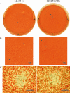
L2(434) and L2(25667R) have similar plaque morphologies. McCoy cells grown in six-well plates were infected with L2(434) or L2(25667R) at approximately 200 IFU/well and neutral red stained for plaques at day 7. (A) Photograph of plaques taken from single wells of a six-well plate. (B) Macroscopic images of L2(434) and L2(25667R) plaques. (C) Microscopic images of L2(434) and L2(25667R) plaques.
FIG. 3.
FIG. 3.
L2(434) but not L2(25667R) inclusions stain positive for glycogen. McCoy cells were infected with L2(434) or L2(25667R) at an MOI of 0.3, fixed at 40 h p.i., and stained for MOMP. The same wells were subsequently stained for glycogen using the iodine staining technique. White arrows in the top panels indicate MOMP staining of individual inclusions of L2(434) (A) and L2(25667R) (A′), while black arrows in the bottom panels (B and B′) identify the same inclusions stained with iodine.
FIG. 4.
FIG. 4.
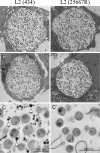
Glycogen localizes to the lumen of mature L2(434) inclusions. Thin sections of L2(434) and L2(25667R) inclusions at 40 h p.i. are shown. (A and A′) Electron-dense extracellular material is present in the L2(434) (A) inclusion but not the L2(25667R) (A′) inclusion. (B and B′) Silver proteinate staining confirms that the electron-dense staining present in the L2(434) inclusion lumen (B) is glycogen, which is absent in the L2(25667R) inclusion (B′). (C and C′) Localization of silver on extracellular material by silver proteinate staining (arrows) indicates the presence of glycogen only in the lumen of the L2(434) (C) and not the L2(25667R) (C′) inclusions.
FIG. 5.
FIG. 5.
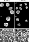
Mature L2(434) and L2(25667R) inclusions are morphologically distinct by phase microscopy. (A to B′) Methanol-fixed inclusions following staining of McCoy cells for MOMP (A and A′) or LPS (B and B′) at 40 h p.i. (magnification, ×600). (C and C′) McCoy cell monolayers were infected at an MOI of 0.3 and viewed live after 40 h p.i. (magnification, ×400). Arrows indicate mature inclusions.
FIG. 6.
FIG. 6.
L2(25667R) exhibits a decreased duration of infection compared to that of L2(434) in a murine model of infection. Shown are mean recoverable IFU values for culture-positive female C3H/HeJ mice challenged with 107 IFU of strain L2(434) or L2(25667R) as shown in Table 3. The actual mean recoverable IFU values for L2(25667R) on days 3 and 7 are 68 and 60, respectively. Error bars represent standard errors of the means.
FIG. 7.
FIG. 7.
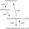
Reaction scheme for glycogen metabolism in C. trachomatis.
FIG. 8.
FIG. 8.
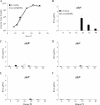
qRT-PCR of glycogen metabolic genes shows a significant difference in glgA expression between L2(434) and L2(25667R). McCoy cells were infected with L2(434) or L2(25667R) (MOI of 1.0) and harvested for RNA and DNA at various times p.i. (PI) (A) Genome copy number was determined using an rpoB-specific primer/probe set and TaqMan quantitative PCR. The experiment was performed twice in triplicate. (B to F) Transcript copy number was determined by TaqMan qRT-PCR and normalized to genome copy number (rpoB) in matched DNA samples. gDNA, genomic DNA.
FIG. 9.
FIG. 9.
Microarray transcriptional analysis of strains L2(434) and L2(25667R) 24 h p.i. (A) Principal-component analysis (PCA) of quantile normalized data. Each spot represents all C. trachomatis data produced for a single chip, where grouping and separation of replicates and conditions, respectively, are demonstrated [blue, L2(25667R); red, L2(434); yellow, mock). (B) Artificial array image constructed in GeneSpring where each spot represents the differential expression of each chlamydial gene, presented in chromosomal order for all 959 ORF represented on the chip. The vertical scale represents changes in transcript levels of L2(25667R) compared to transcript levels of L2(434). (C) Venn diagram identifying the 29 gene transcripts that passed the statistical and quality tests performed as described in Materials and Methods (signal, signal above background; call, call consistency; 2×, twofold or higher minimum threshold filter; SAM, significance analysis of microarrays; ttest, Student's t test). (D) Chromosomal order of the 29 genes identified above (C).

References

    1. Albert, T. J., D. Dailidiene, G. Dailide, J. E. Norton, A. Kalia, T. A. Richmond, M. Molla, J. Singh, R. D. Green, and D. E. Berg. 2005. Mutation discovery in bacterial genomes: metronidazole resistance in Helicobacter pylori. Nat. Methods 2951-953. - PubMed
    1. An, Q., G. Radcliffe, R. Vassallo, D. Buxton, W. J. O'Brien, D. A. Pelletier, W. G. Weisburg, J. D. Klinger, and D. M. Olive. 1992. Infection with a plasmid-free variant chlamydia related to Chlamydia trachomatis identified by using multiple assays for nucleic acid detection. J. Clin. Microbiol. 302814-2821. - PMC - PubMed
    1. Azuma, Y., H. Hirakawa, A. Yamashita, Y. Cai, M. A. Rahman, H. Suzuki, S. Mitaku, H. Toh, S. Goto, T. Murakami, K. Sugi, H. Hayashi, H. Fukushi, M. Hattori, S. Kuhara, and M. Shirai. 2006. Genome sequence of the cat pathogen, Chlamydophilia felis. DNA Res. 1315-23. - PubMed
    1. Bai, G., E. Smith, A. Golubov, J. Pata, and K. A. McDonough. 2007. Differential gene regulation in Yersinia pestis pseudotuberculosis: effects of hypoxia and potential role of a plasmid regulator. Adv. Exp. Med. Biol. 603131-144. - PubMed
    1. Belland, R. J., G. Zhong, D. D. Crane, D. Hogan, D. Sturdevant, J. Sharma, W. L. Beatty, and H. D. Caldwell. 2003. Genomic transcriptional profiling of the developmental cycle of Chlamydia trachomatis. Proc. Natl. Acad. Sci. USA 1008478-8483. - PMC - PubMed

Publication types

MeSH terms

Substances

Associated data