. 2008 May;28(10):3401-9.
doi: 10.1128/MCB.00006-08. Epub 2008 Mar 17.

Transcription-coupled methylation of histone H3 at lysine 36 regulates dosage compensation by enhancing recruitment of the MSL complex in Drosophila melanogaster

Affiliations

Transcription-coupled methylation of histone H3 at lysine 36 regulates dosage compensation by enhancing recruitment of the MSL complex in Drosophila melanogaster

Oliver Bell et al. Mol Cell Biol. 2008 May.

Abstract

In Drosophila melanogaster, dosage compensation relies on the targeting of the male-specific lethal (MSL) complex to hundreds of sites along the male X chromosome. Transcription-coupled methylation of histone H3 lysine 36 is enriched toward the 3' end of active genes, similar to the MSL proteins. Here, we have studied the link between histone H3 methylation and MSL complex targeting using RNA interference and chromatin immunoprecipitation. We show that trimethylation of histone H3 at lysine 36 (H3K36me3) relies on the histone methyltransferase Hypb and is localized promoter distal at dosage-compensated genes, similar to active genes on autosomes. However, H3K36me3 has an X-specific function, as reduction specifically decreases acetylation of histone H4 lysine 16 on the male X chromosome. This hypoacetylation is caused by compromised MSL binding and results in a failure to increase expression twofold. Thus, H3K36me3 marks the body of all active genes yet is utilized in a chromosome-specific manner to enhance histone acetylation at sites of dosage compensation.


Figures

FIG. 1.
FIG. 1.
High-resolution analysis of di- and trimethylation of H3K36 on autosomal and dosage-compensated genes. ChIP analysis of Drosophila SL2 cells using antibodies specific for H3K36me2 or H3K36me3 and quantification by real-time PCR. Shown are the average and standard deviation of ChIP enrichments from at least three independent experiments normalized to histone H3 occupancy. The x axis reflects the base pair position relative to the transcriptional start site. The y axis reflects enrichment (bound/input values are displayed as percent recovery of input DNA). H3K36me2, left scale; H3K36me3, right scale. Numbers in graphs are gene identification numbers according to Flybase.
FIG. 2.
FIG. 2.
Hypb binds autosomes and the male X chromosome. Polytene chromosomes of male third-instar larvae were stained with antibodies against Hypb (green) and MSL1 (red). DNA was visualized by Hoechst staining (blue). (A) Hypb localized preferentially to euchromatic interband regions, suggesting a general role in transcription. (B) Hypb partially localizes to sites of MSL1 enrichment along the X chromosome, consistent with a role in the trimethylation of H3K36 on dosage-compensated genes.
FIG. 3.
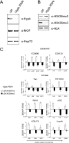
FIG. 3.
RNAi knockdown of Hypb has similar effects at autosomal and X-linked loci. (A) Western blot analysis using antibody specific for Hypb reveals efficient reduction of Hypb in male Drosophila SL2 cells. Hsp70 and MOF remain unaffected by RNAi knockdown and serve as loading controls. (B) Reduction of Hypb results in a reduction of H3K36me3 and a coinciding increase of H3K36me2. H2A serves as a loading control. (C) Levels of H3K36 methylation states in RNAi and control cells were compared by ChIP followed by real-time PCR analysis. Shown is the ratio of H3K36me enrichments (change [n-fold], y axis) of RNAi over control cells relative to the position from the transcription start site (x axis). Effects on H3K36 methylation states at individual loci reflect bulk changes upon Hypb knockdown.
FIG. 4.
FIG. 4.
H3K36 trimethylation is required for H4K16 hyperacetylation of the dosage-compensated X chromosome. (A) Hypb RNAi results in a reduction of bulk H4K16ac in male SL2 cells as indicated by Western blot analysis. (B) Comparison of changes in H4K16ac along autosomal and dosage-compensated X-linked genes upon RNAi by ChIP and real-time PCR. At autosomal genes, H4K16ac levels increase upon reduction of Hypb, whereas levels at dosage-compensated genes decrease.
FIG. 5.
FIG. 5.
H3K36 trimethylation enhances binding of the MSL complex and transcriptional upregulation at dosage-compensated genes. (A) MOF and MSL1 enrichments at dosage-compensated genes were compared with untreated and Hypb RNAi cells by ChIP and real-time PCR analysis. In control cells, both proteins displayed a similar pattern, localizing preferentially in the 3′ end of genes. Hypb knockdown led to diminished levels of MOF and MSL1 at all positions along Par-6, CG8173, and Ucp4A. MOF was also depleted from high-affinity sites along the roX2 gene, while the level of MSL1 remained largely unaffected. (B) Relative mRNA expression from dosage-compensated and noncompensated genes after Hypb knockdown. Displayed are average mRNA levels of five independent experiments, normalized to a mitochondrial RNA, comparing untreated and Hypb knockdown cells by quantitative real-time PCR (14). Upon Hypb RNAi, mRNA expression levels of 11 dosage-compensated genes are reduced approximately twofold. Expression levels of noncompensated X-linked runt as well as autosomal spt4 remain unchanged.
FIG. 6.
FIG. 6.
Model for MSL complex targeting to sites of dosage compensation in Drosophila. Genes along the X chromosome have various affinities for MSL complex binding. High-affinity sites (red) can attract partially assembled MSL complexes independent of transcription-coupled chromatin modifications. However, H3K36me3 is still necessary to facilitate robust interaction with MOF and MSL3. In comparison, the majority of target genes contain promoter-distal sequence elements, which have relatively weak affinity for MSL recruitment (low-affinity sites [yellow]). At these genes, transcription-dependent H3K36me3 (blue arch represents concentration of H3K36me3) enhances recognition and stable binding of fully assembled MSL complexes. Thus, robust recruitment of the MSL proteins to the dosage-compensated X chromosome relies on combined contributions of degenerate sequence elements and transcription-coupled histone modifications.

References

    1. Akhtar, A., and P. B. Becker. 2000. Activation of transcription through histone H4 acetylation by MOF, an acetyltransferase essential for dosage compensation in Drosophila. Mol. Cell 5367-375. - PubMed
    1. Alekseyenko, A. A., E. Larschan, W. R. Lai, P. J. Park, and M. I. Kuroda. 2006. High-resolution ChIP-chip analysis reveals that the Drosophila MSL complex selectively identifies active genes on the male X chromosome. Genes Dev. 20848-857. - PMC - PubMed
    1. Barski, A., S. Cuddapah, K. Cui, T. Y. Roh, D. E. Schones, Z. Wang, G. Wei, I. Chepelev, and K. Zhao. 2007. High-resolution profiling of histone methylations in the human genome. Cell 129823-837. - PubMed
    1. Bell, O., C. Wirbelauer, M. Hild, A. N. Scharf, M. Schwaiger, D. M. MacAlpine, F. Zilbermann, F. van Leeuwen, S. P. Bell, A. Imhof, D. Garza, A. H. Peters, and D. Schubeler. 2007. Localized H3K36 methylation states define histone H4K16 acetylation during transcriptional elongation in Drosophila. EMBO J. 264974-4984. - PMC - PubMed
    1. Carrozza, M. J., B. Li, L. Florens, T. Suganuma, S. K. Swanson, K. K. Lee, W. J. Shia, S. Anderson, J. Yates, M. P. Washburn, and J. L. Workman. 2005. Histone H3 methylation by Set2 directs deacetylation of coding regions by Rpd3S to suppress spurious intragenic transcription. Cell 123581-592. - PubMed

Publication types

MeSH terms

LinkOut - more resources