. 2008 Jun;56(6):587-95.
doi: 10.1369/jhc.2008.950675. Epub 2008 Mar 17.

Generation and characterization of Ptf1a antiserum and localization of Ptf1a in relation to Nkx6.1 and Pdx1 during the earliest stages of mouse pancreas development

Affiliations

Generation and characterization of Ptf1a antiserum and localization of Ptf1a in relation to Nkx6.1 and Pdx1 during the earliest stages of mouse pancreas development

Jacob Hald et al. J Histochem Cytochem. 2008 Jun.

Abstract

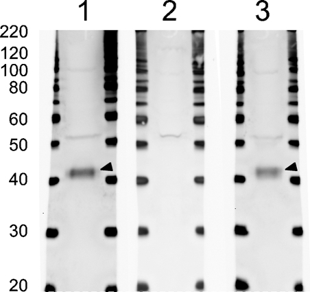
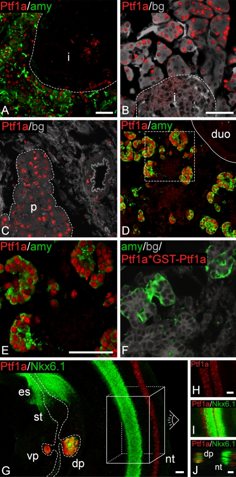
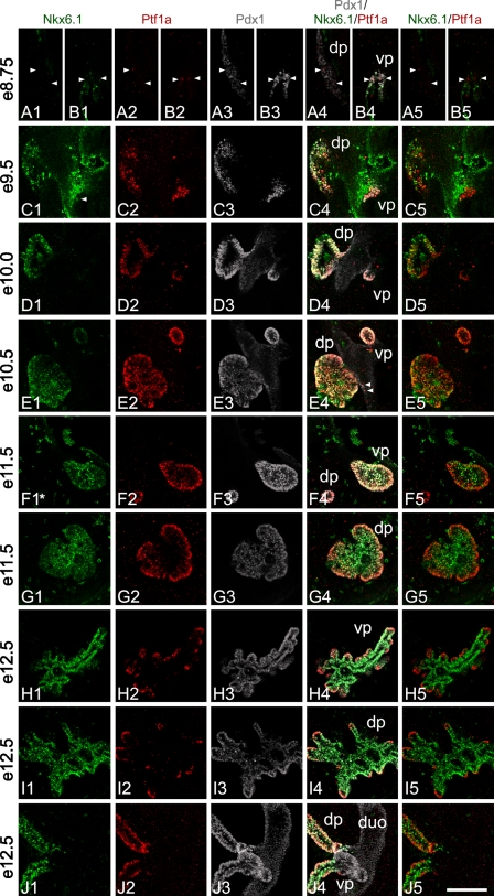
Ptf1a and Pdx1 are critical transcription factors of early pancreatic development, as shown by loss of function studies where lack of each gene alone causes almost complete pancreas agenesis. Ptf1a is particularly interesting because it is linked to a recently reported signature gene expression profile associated with the multipotent condition. Few useful antibody reagents have been available for consistent and reliable immunohistochemical visualization of Ptf1a protein expression in the early developing pancreas in which the level of production of this critical regulator seems to be very low. We describe a novel rabbit antibody raised against the c-terminal portion of the mouse Ptf1a protein and report immunodetection, for the first time, as early as embryonic day (e) 8.5-e8.75 in the dorsal and ventral buds of the mouse pancreas as well as in the neural tube at e10.0. Detailed confocal analysis identifies an abundant triple-positive (Ptf1a(+)/Nkx6.1(+)/Pdx1(+)) putative early multipotent pancreatic progenitor cell that marks the e9.5 dorsal pancreas and e10.5 ventral pancreas. Furthermore, expression patterns of Nkx6.1 vs Ptf1a subsequently segregate during branching morphogenesis (trunk vs tip), ending up marking two distinct cell populations of progenitors at e12.5. From e15.5 (mouse) and in adult pancreas (mouse, rat, and human), the Ptf1a antibody marks only acinar cell nuclei, as expected for its subsequent role in committing/maintaining cells in this differentiated state. In summary, this antibody is a novel tool to further characterize important early steps of pancreas differentiation. This manuscript contains online supplemental material at http://www.jhc.org. Please visit this article online to view these materials.


Figures

Figure 1
Figure 1
Anti-Ptf1a serum recognizes a 42-kDa protein. Whole cell lysate from embryonic day (e) 15.5 pancreas was loaded in Lanes 1, 2, and 3. In Lane 1, the Ptf1a antiserum recognizes a weak band (∼52 kDa) and a strong band (arrowhead; ∼42 kDa) correlating well with the calculated molecular mass of 37.7 kDa of Ptf1a. In Lane 2, Ptf1a antiserum was preincubated with glutathione-S-transferase (GST)–Ptf1a and the intense 42-kDa band is not detected, whereas the weak 52-kDa band is still present. In Lane 3, the Ptf1a antiserum was preincubated with an unspecific control, GST–Nkx6.1, which does not interfere with the staining pattern as expected.
Figure 2
Figure 2
Ptf1a antiserum stains specifically in the pancreas and neural tube. (A) Double staining for Ptf1a and amylase on adult mouse cryosection. Robust immunoreactivity is observed for Ptf1a. Immunoreactivity is localized to the nucleus of amylase-positive cells and not in the islet outlined by the broken line. (B) Staining for Ptf1a on adult rat cryosection. Intense immunoreactivity for Ptf1a is evident in the nucleus of acinar cells of the pancreas. A negative islet is outlined with a broken line. (C) Ptf1a staining of adult human paraffin section. Ptf1a immunoreactivity marks acinar cells of the pancreas marked by the broken line. (D,E) Cryosection of e15.5 mouse section stained for Pft1a and amylase. Area marked by the broken line in D is shown in E. Full line marks the pancreas–duodenum boundary. In the gut region, Ptf1a-like immunoreactivity is observed only in the pancreas and is localized to the nucleus of the developing acinar cells. As observed in E, few Ptf1a-positive cells are negative for amylase at this time point. (F) Ptf1a antiserum preincubated with GST–Ptf1a (Ptf1a*GST–Ptf1a) prior to staining of an e15.5 mouse cryosection. Preincubating Ptf1a antiserum with GST–Ptf1a results in lack of Ptf1a immunoreactivity, compare with E. (G–I) Whole-mount immunofluorescence (IF) was performed on e10.5 mouse embryos for Ptf1a and Nkx6.1. (G) Ptf1a-like immunoreactivity is observed only in the pancreas and neural tube. In the pancreas, Ptf1a staining colocalizes with Nkx6.1 immunoreactivity. In the neural tube, Ptf1a reactivity is localized more dorsal than Nkx6.1 reactivity. Data in H and I are recorded looking from the dorsal side of an embryo down on the neural tube represented by the box and eye drawn in G. The more dorsal expression of Ptf1a compared with Nkx6.1 is also evident on the orthogonal section in J. Nkx6.1- and Ptf1a-like immunoreactivities are observed in both sides of the neural tube (H–J). Whole-mount double staining was done for Ptf1a and Nkx6.1 and stacks of optical sections recorded on a confocal microscope. Pictures shown in G–I represent two or more superimposed optical sections. This is done to present 3D information in a 2D format; thus, cells that appear to be overlapping are not necessarily so. (B,C,F) Gray/background is autofluorescence from cells to outline the tissue. i, islet; duo, duodenum; dp, dorsal pancreas; vp, ventral pancreas; nt, neural tube; st, stomach; es, esophagus. Identical magnifications for A and D, B and C, E and F, H and I. Bar = 50 μm.
Figure 3
Figure 3
Ptf1a, Nkx6.1, and Pdx1 localization in the developing pancreas. Whole-mount triple staining was done for Ptf1a, Nkx6.1, and Pdx1 from e8.5 to e12.5 mouse embryos. Stacks of optical sections were recorded on a confocal microscope. Pictures shown in rows A,B,F,G,H,I are single optical sections from a stack, whereas pictures in rows C,D,E,J are composed of several cropped optical sections. This is done to present 3D information in a 2D way avoiding, at the same time, superimposing cells not in the same optical section. All optical sections can be viewed on supplementary movie SM1A–J. Asterisk in F1 indicates that the original Nkx6.1 signal has been bleached by the laser, whereas recording the dorsal pancreas seen in row G thus represents an artifact. (A1–5, B1–5) At ∼e8.75, the first weak Ptf1a-positive cells can be observed in the dorsal pancreas (arrowheads in A1–5). In the ventral pancreas (B1–5), immunoreactivity observed in scattered cells is stronger compared with positive cells in the dorsal pancreas. The first cells positive for Ptf1a appear at ∼e8.5 in the ventral pancreas (data not shown). The dorsal pancreas is devoid of Nkx6.1-positive cells (A1) and Ptf1a expression thus precedes that of Nkx6.1 in the dorsal pancreas. (C1–5) At e9.5, robust Ptf1a expression is observed in the dorsal and ventral pancreas. In the ventral pancreas, Ptf1a is expressed at levels comparable to what is observed later in development. Nkx6.1 is readily observed in the dorsal pancreas. In the ventral pancreas only very few and low Nkx6.1-expressing cells are observed (arrowhead) (see also supplemental movie SM1C). Thus, most Ptf1a-positive cells of the ventral pancreas are negative for Nkx6.1, whereas in the dorsal pancreas most Ptf1a-positive cells are also positive for Nkx6.1. In the dorsal pancreas, Ptf1a- and Nkx6.1-positive cells are found in what appears to be a disorganized pattern, whereas Ptf1a expression pattern in the ventral pancreas is much more uniform. (D1–5) At e10.0, cells positive for Ptf1 and Nkx6.1 are distributed in what appears to be a more organized epithelium compared with e9.5. In the dorsal pancreas, intense Ptf1 and Nkx6.1 immunoreactivity is observed at a level that is comparable to what is seen later in development. Ptf1a and Nkx6.1 positive cells are generally distributed in the bud in a random pattern however, clusters of single positive cells are also present. Nearest to the duodenum, cells tend to be more Ptf1a positive and Nkx6.1 negative/low compared with the rest of the bud, and in some bud areas cells that are Nkx6.1 positive and Ptf1a weak/negative are present. In the ventral pancreas, Ptf1a continues to be expressed at high levels, whereas Nkx6.1 is close to undetectable. (E1–5) At e10.5, Ptf1a, Pdx1, and Nkx6.1 are close to uniformly expressed in the ventral bud and to a large degree in the dorsal bud. Nkx6.1 immunoreactivity reappears in the ventral bud although immunoreactivity is weak. Ptf1a is strongly expressed in both buds. In the dorsal pancreas the tendency of having Ptf1a-positive, Nkx6.1-negative cells nearest to the duodenum continues. Clusters of cells negative for Ptf1a form holes in the otherwise uniform optical sections of Ptf1a-expressing dorsal bud cells. These cells are Nkx6.1 positive. Similar clusters are also observed budding off the Ptf1a-positive area. Such clusters are likely forming endocrine cells. In the rim of the bud, Pdx1 immunoreactivity is more intense compared with the other cells (E3). Furthermore, there is a small tendency for the very center of the dorsal bud to be more Nkx6.1 positive than Ptf1a positive. A few Ptf1a-positive cells can be observed in the duodenum outside the bud area (arrowheads in E4). (F1–5, G1–5) At e11.5, in the ventral pancreas Ptf1a, Nkx6.1, and Pdx1 are coexpressed in almost all cells. Pdx1 is expressed at similar intense levels throughout the bud, but Ptf1a and Nkx6.1 start to display opposite expression levels. This results in a cell population in the rim of the bud that expresses Ptf1a at elevated levels and coexpresses Nkx6.1 weakly, and cells in the center that express Ptf1a weakly and coexpress Nkx6.1 at an elevated level. Expression in the dorsal pancreas is like that of the ventral, just more distinct as Ptf1a expression is extinguished in the very center of the bud, but Nkx6.1 continues to be expressed weakly in the rim cells. In the forming stalk of the dorsal pancreas, Ptf1a and Pdx1 are strongly expressed, whereas Nkx6.1 expression is weaker. (H1–5, I1–5) At e12.5, expression levels and localization of Ptf1a and Nkx6.1 are almost reciprocal, whereas Pdx1 remains more uniformly expressed. Ptf1a is mainly localized to the tips of the branching pancreas where it is expressed at high levels. Nkx6.1 is mainly localized to all cells but the tip cells and the tip cells that do express Nkx6.1 do so at low levels. A few scattered cells express Pdx1 at an elevated level. Such cells are likely forming insulin cells. (J1–5) At e12.5, in the stalk regions Ptf1a-positive cells are localized to the rim of the stalks. Nkx6.1-positive cells are localized throughout the stalk with the weak-expressing cells in the rim and strong-expressing cells in the center. Pdx1 is uniformly localized and expressed. dp, dorsal pancreas; vp, ventral pancreas; duo, duodenum. Magnification is the same in all pictures. Bar = 100 μm.

References

    1. Adell T, Gomez-Cuadrado A, Skoudy A, Pettengill OS, Longnecker DS, Real FX (2000) Role of the basic helix-loop-helix transcription factor p48 in the differentiation phenotype of exocrine pancreas cancer cells. Cell Growth Differ 11:137–147 - PubMed
    1. Afelik S, Chen Y, Pieler T (2006) Combined ectopic expression of Pdx1 and Ptf1a/p48 results in the stable conversion of posterior endoderm into endocrine and exocrine pancreatic tissue. Genes Dev 20:1441–1446 - PMC - PubMed
    1. Ahnfelt-Ronne J, Jorgensen MC, Hald J, Madsen OD, Serup P, Hecksher-Sorensen J (2007) An improved method for 3D reconstruction of protein expression patterns in intact mouse and chicken embryos and organs. J Histochem Cytochem 55:925–930. Published online May 3, 2007. 10.1369/jhc.7A7226 - PubMed
    1. Beres TM, Masui T, Swift GH, Shi L, Henke RM, MacDonald RJ (2006) PTF1 is an organ-specific and Notch-independent basic helix-loop-helix complex containing the mammalian Suppressor of Hairless (RBP-J) or its paralogue, RBP-L. Mol Cell Biol 26:117–130 - PMC - PubMed
    1. Candia AF, Wright CV (1996) Differential localization of Mox-1 and Mox-2 proteins indicates distinct roles during development. Int J Dev Biol 40:1179–1184 - PubMed

Publication types

MeSH terms