. 2008 Mar 15;22(6):810-23.
doi: 10.1101/gad.462608.

Auxin influx carriers stabilize phyllotactic patterning

Affiliations

Auxin influx carriers stabilize phyllotactic patterning

Katherine Bainbridge et al. Genes Dev. .

Abstract

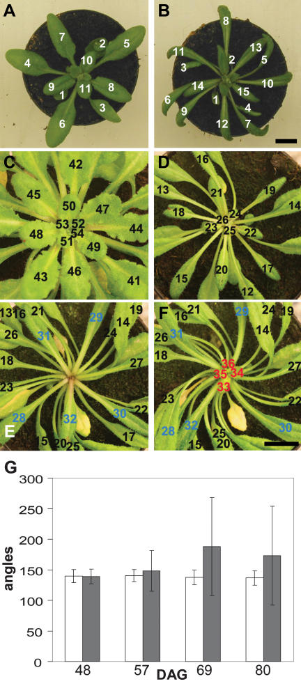
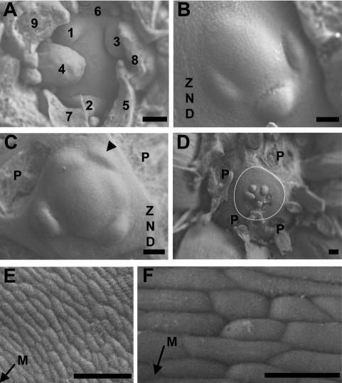
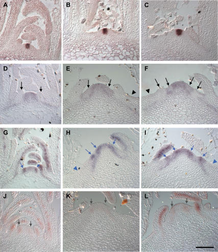
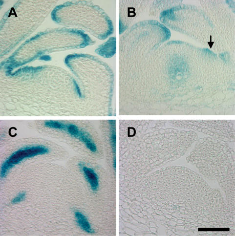
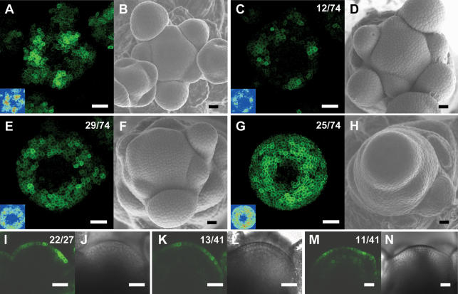
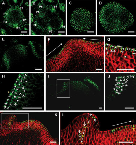
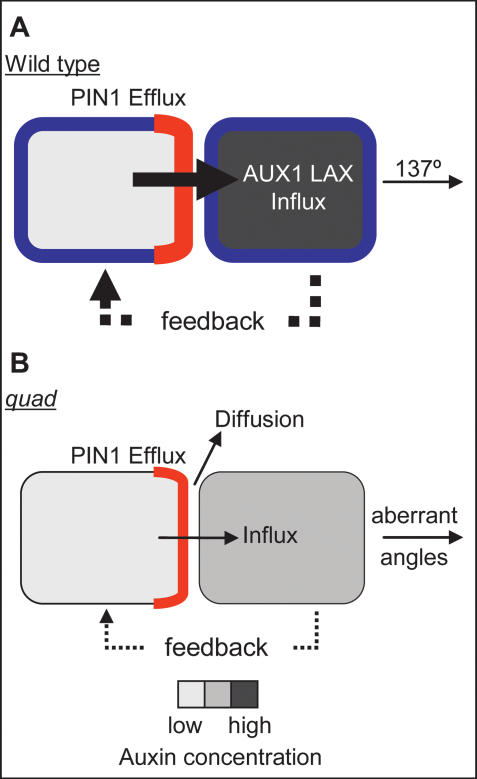
One of the most striking features of plant architecture is the regular arrangement of leaves and flowers around the stem, known as phyllotaxis. Peaks in concentration of the plant hormone auxin, generated by the polar localization of the PIN1 auxin efflux carrier, provide the instructive signal for primordium initiation. This mechanism generates the spacing between neighboring primordia, which results in regular phyllotaxis. Studies of the role of auxin transport in phyllotactic patterning have focused on PIN1-mediated efflux. Recent computer simulations indicate an additional role for transporter-mediated auxin uptake. Mutations in the AUX1 auxin influx carrier have not, however, been reported to cause an aerial phenotype. Here, we study the role of AUX1 and its paralogs LAX1, LAX2, and LAX3. Analysis of the quadruple mutant reveals irregular divergence angles between successive primordia. A highly unusual aspect of the phenotype is the occurrence of clusters of primordia, in violation of classical theory. At the molecular level, the sharp peaks in auxin levels and coordinated PIN polarization are reduced or lost. In addition, the increased penetrance of the phenotype under short-day conditions suggests that the AUX LAX transporters act to buffer the PIN-mediated patterning mechanism against environmental or developmental influences.


Figures

Figure 1.
Figure 1.
Leaf initiation in wild-type and quadruple aux1 lax mutant plants. Photographs showing vegetative rosettes of wild-type and quad plants under long- and short-day conditions. (A) Wild type; long days. (B) quad; long days showing narrow, twisted leaf blades and regular, spiral phyllotaxis. (C) Wild type; short days, 73 DAG. (DF) The same short-day-grown quad plant at three different time points. (D) quad 62 DAG. (E) quad 73 DAG. (F) quad 78 DAG. Leaves numbered in order of initiation. (Blue) Leaves initiated immediately before arrest; (red) leaves initiated after arrest. Bars, 1 cm. (G) Graph showing the mean divergence angle between successively formed leaves at four different time points. (White bars) quad; (gray bars) wild type. Error bars represent the standard error of the mean. n = 20.
Figure 2.
Figure 2.
Irregular primordium initiation at vegetative meristems of the quadruple aux1 lax mutant. Transversal scanning electron microscope images of wild-type and quad meristems. (A) Wild type. (BF) quad. Leaf primordia are labeled in the order of initiation, with 1 denoting the youngest visible primordium. In cases where the order of initiation is unclear, primordia are not numbered. P denotes the base of primordia removed during dissection. The arrowhead in C indicates an abortive primordium that has initiated and moved away from the meristem without developing further. The white line in D outlines the periphery of the ZND. (E,F) Cells of the ZND. The arrows indicate the direction of the meristem (M). Bars: A–C,E, 50 μm; D, 100 μm; F, 20 μm. Plants dissected at 55 DAG.
Figure 3.
Figure 3.
In situ analysis of expression of meristem and early primordium genes in wild type and the quadruple aux1 lax mutant. Central sections of a series of longitudinal sections through wild-type and quad vegetative meristems. (A) CLV3 in wild type. (B,C) CLV3 in quad. (D) STM in wild type. (E,F) STM in quad. (G) ANT in wild type. (H,I) ANT in quad. (J) MP in wild type. (K,L) MP in quad. Black arrows indicate sites of STM down-regulation in incipient or bulging primordia. Black arrowheads indicate lack of STM expression in the outer ZND. Blue arrows indicate sites of ANT up-regulation in incipient or bulging primordia. Blue arrowheads indicate lack of ANT expression in the outer ZND. Gray arrows indicate MP expression in the peripheral zone of the meristem. (A,B,D,G,J) 33–39 DAG, (C,E,F,H,I,K,L) 55–65 DAG. Bars, 50 μm.
Figure 4.
Figure 4.
Expression of AUX1 and LAX promoter∷GUS at vegetative meristems. Central, longitudinal sections through vegetative meristems in the wild-type background. (A) AUX1pr∷GUS. (B) LAX1pr∷GUS. Arrow indicates expression in the peripheral zone at the site of a bulging primordium. (C) LAX2pr∷GUS. (D) LAX3pr∷GUS, 40 DAG. Histological GUS staining. Bars, 50 μm.
Figure 5.
Figure 5.
The pattern of DR5∷GFP expression is altered in aux1 lax1 lax2 triple mutant inflorescence meristems. (A) Maximal projection of transversal confocal scans of a wild-type meristem expressing DR5∷GFP. (Inset) LUT signal intensity monitor. (Blue) Low intensity; (red) high intensity. (B) Scanning electron microscope image of the same wild-type meristem as in A. (C,E,G) Maximal projections of transversal confocal scans of DR5∷GFP-expressing aux1 lax1 lax2 triple mutant meristems displaying increasingly severe phenotypes. (Inset) LUT signal intensity monitor. Representative meristems showing smaller and weaker peaks (C), broader and less defined peaks (E), or expression throughout the peripheral zone (G). (D,F,H) Scanning electron microscope images of the same meristems as in C, E, and G, respectively. (I) Longitudinal confocal section through a DR5∷GFP-expressing wild-type meristem showing no GFP expression in inner layers, except below initiation sites. (J) Transmission light picture of the wild-type meristem shown in I. (K,M) Longitudinal confocal section through two aux1 lax1 lax2 triple mutant meristems, showing very weak (K) or weak (M) DR5∷GFP expression in inner layers. (L,N) Transmission light pictures of the meristems shown in K and M. Bars, 25 μm. Numbers indicate the number of occurrences over the total number of observations.
Figure 6.
Figure 6.
PIN1 polarization in the quadruple aux1 lax mutant. Green signal is PIN1 protein. Red signal is calcafluor staining of the cell walls. PIN1 localization visualized in maximal projections of serial optical sections, made by confocal imaging. (AD) Transversal sections. (A,B) Wild-type meristem. Primordia labeled in order of initiation, with P1 the most recently initiated. (A) Ten to twenty microns below the meristem summit, showing convergent PIN1 polarization in incipient and outgrowing primordia in the L1 of the peripheral zone. (B) Twenty to thirty microns below the summit with PIN1 polarization toward developing vasculature of incipient and outgrowing primordia. (C,D) quad meristem. (C) Ten to twenty microns below the quad meristem summit, showing diffuse PIN1 polarization. (D) Twenty to thirty microns below the summit, showing diffuse PIN1 polarization. (EL) Longitudinal sections. (E) Central section through a wild-type meristem, showing convergent PIN1 localization in incipient and outgrowing primordia and basipetal PIN1 polarization along the sites of developing vasculature. (F) Central section through a quad meristem with no recently initiated primordia, showing polarization of PIN1 toward the meristem summit in the L1 layer stretching to the periphery of the ZND. (G) Enlargement of the right-hand ZND of the meristem shown in F, showing polarization of PIN1 toward the meristem summit in the L1 and apical polarization in the L2. (H) Peripheral zone of a quad meristem with no newly initiated primordia showing convergent polarization in two cells of the PZ (yellow star). (I) A quad meristem with an incipient primordium in the PZ on the left and a recently initiated primordium to the right. (J) Enlargement of the incipient primordium indicated by the rectangle in I, showing an L1 convergence point (white star) and basal polarization of PIN1 in underlying cell layers. (K) A quad meristem with a newly initiated primordium in the PZ on the left. (L) Enlargement of the newly initiated primordium indicated by the rectangle in K, showing basal polarization of PIN1. Closed-headed arrows show the general direction of polarization in a group of cells. Open-headed arrows show the direction of PIN1 polarization in each cell. White rectangles indicate areas enlarged in the next panel. Bars, 25 μm.
Figure 7.
Figure 7.
Model for the role of AUX1 LAX importers in phyllotaxis. (A) Two wild-type cells. (B) Two aux1 lax mutant cells. PIN1 (red), AUX1 LAX (blue), and auxin concentration (gray) are represented. In both wild-type and quad, PIN1 polarizes toward the neighboring cell with the higher auxin concentration and exports auxin from the cell. (A) In wild type, auxin is rapidly taken up into the next cell due to AUX1 LAX function, causing an increase in auxin concentration in this cell. The higher auxin concentration in this cell feeds back to control PIN1 polarity. This causes further efflux toward this cell, which is rapidly taken up and further increases auxin concentration. This tightly controlled generation of peaks and troughs in auxin concentration generates a regular angle of 137° between successive primordia. (B) In aux1 lax mutants, auxin leaves the cell via PIN1-mediated efflux. The absence of AUX1 LAX reduces the efficiency with which auxin is taken up into the next cell, and therefore more diffusion of auxin can occur in the apoplastic space. Some auxin may enter the next cell to increase the auxin concentration of the cell. Any increase in auxin concentration would feed back to control PIN1 polarity, increasing efflux and further increasing auxin concentration in the next cell. This less regulated generation of auxin peaks results in irregular spacing between primordia. In addition, peaks above the threshold for primordium initiation, but below the threshold level for full primordium differentiation, would result in the abortive primordia seen in the quad.

References

    1. Abas L., Benjamins R., Malenica N., Paciorek T., Wirniewska J., Moulinier-Anzola J.C., Sieberer T., Friml J., Luschnig C. Intracellular trafficking and proteolysis of the Arabidopsis auxin-efflux facilitator PIN2 are involved in root gravitropism. Nat. Cell Biol. 2006;8:249–256. - PubMed
    1. Aida M., Vernoux T., Furutani M., Traas J., Tasaka M. Roles of PIN-FORMED1 and MONOPTEROS in pattern formation of the apical region of the Arabidopsis embryo. Development. 2002;129:3965–3974. - PubMed
    1. Benkova E., Michniewicz M., Sauer M., Teichmann T., Jurgens G., Friml J. Local, efflux-dependent auxin gradients as a common module for plant organ formation. Cell. 2003;115:591–602. - PubMed
    1. Bennett M.J., Marchant A., Green H.G., May S.T., Ward S.P., Millner P.A., Walker A.R., Schulz B., Feldmann K.A. Arabidopsis AUX1 gene: A permease-like regulator of root gravitropism. Science. 1996;273:948–950. - PubMed
    1. Carabelli M., Possenti M., Sessa G., Ciolfi A., Sassi M., Morelli G., Ruberti I. Canopy shade causes a rapid and transient arrest in leaf development through auxin-induced cytokinin oxidase activity. Genes & Dev. 2007;21:1863–1868. - PMC - PubMed

Publication types

MeSH terms