. 2008 Mar 25;105(12):4691-6.
doi: 10.1073/pnas.0711531105. Epub 2008 Mar 17.

Myosin V and Kinesin act as tethers to enhance each others' processivity

Affiliations

Myosin V and Kinesin act as tethers to enhance each others' processivity

M Yusuf Ali et al. Proc Natl Acad Sci U S A. .

Abstract

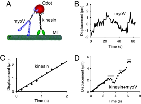
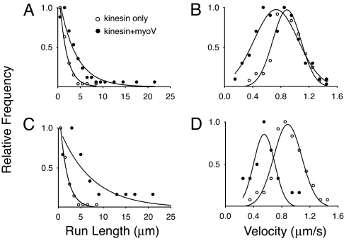
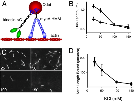
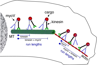
Organelle transport to the periphery of the cell involves coordinated transport between the processive motors kinesin and myosin V. Long-range transport takes place on microtubule tracks, whereas final delivery involves shorter actin-based movements. The concept that motors only function on their appropriate track required further investigation with the recent observation that myosin V undergoes a diffusional search on microtubules. Here we show, using single-molecule techniques, that a functional consequence of myosin V's diffusion on microtubules is a significant enhancement of the processive run length of kinesin when both motors are present on the same cargo. The degree of run length enhancement correlated with the net positive charge in loop 2 of myosin V. On actin, myosin V also undergoes longer processive runs when kinesin is present on the same cargo. The process that causes run length enhancement on both cytoskeletal tracks is electrostatic. We propose that one motor acts as a tether for the other and prevents its diffusion away from the track, thus allowing more steps to be taken before dissociation. The resulting run length enhancement likely contributes to the successful delivery of cargo in the cell.


Conflict of interest statement

The authors declare no conflict of interest.

Figures

Fig. 1.
Fig. 1.
Motor movement on microtubules. (A) Cartoon of the experimental setup. Microtubules (MT) are adhered to a glass coverslip. Full-length constructs of myoV, kinesin, or both motors are attached to carboxylated Qdots (see Methods), and visualized by in an objective-type total internal reflectance microscope. Experiments with constitutively active constructs of myoV and kinesin, attached to streptavidin-Qdots via a C-terminal biotin tag, were also performed. (B) One-dimensional diffusion of myoV on microtubules with no net displacement. (C) Smooth movement routinely observed when kinesin alone is attached to the Qdot. Data obtained at the onset of movement when the Qdot appeared on the microtubule until its disappearance when kinesin terminated its run. The velocity for this kinesin molecule was 1.13 μm/s, estimated from the linear regression. Images were captured at 5 frames/s. (D) New behavior seen when both kinesin and myoV are attached to a Qdot (data are from SI Movie 1). For kinesin/myoV-labeled Qdots, numerous runs were characterized by periods of smooth directed movement, as highlighted by the regression through the first 3 s, interspersed with diffusive searches identified by horizontal lines. The periods of smooth movement were characteristic of the movement seen with Qdots labeled only with kinesin (as in C), having velocities (0.80 μm/s for the first 3 s of this run based on regression) comparable to that of kinesin alone (see Table 1). Data obtained at the onset of movement when the Qdot appeared on the microtubule until its disappearance when the run terminated. Note that the scales are different when comparing the Qdot movement here which has both kinesin and myoV attached to that of a Qdot labeled solely with kinesin in C. Image capture rate of 5 frames/s.
Fig. 2.
Fig. 2.
Run length (A and C) and velocity (B and D) distributions on microtubules. (A and B) Data obtained when Qdots were incubated with both kinesin and myoV (filled circles), or with kinesin alone (open circles). (C and D) Analysis of the subpopulation of runs (22% of total) that included obvious diffusive events or pauses. The run length and velocity of the entire run (filled circles), are compared with the run length and velocity of kinesin alone (open circles, as in A and B). The relative frequencies were determined by using the total number of runs for a given experimental condition as reported in Table 1.
Fig. 3.
Fig. 3.
Processive movement on actin filaments. (A) Cartoon of the experimental setup. Actin filaments are adhered to a glass coverslip. Constitutively active constructs of myoV-HMM and kinesin-ΔC, both of which contain a biotinated tag at their C terminus, are attached to streptavidin-Qdots (see Methods), and visualized by in an objective-type total internal reflectance microscope. (B) Run lengths as a function of KCl concentration for myoV-HMM alone (open triangles) and for myoV in the presence of kinesin-ΔC (filled triangles). Run lengths at both 25 and 50 mM KCl in the presence of kinesin were statistically different (P < 0.05) from those in the absence of kinesin, using the Kolmogorov-Smirnov Test. (C) Raw images obtained of actin bound to a kinesin-coated glass coverslip at varying KCl concentration (concentration in mM KCl in lower left corner). (D) Average total length of actin bound per 8,700 μm2 of a kinesin-coated glass coverslip, as a function of KCl concentration.
Fig. 4.
Fig. 4.
Model illustrating how myoV and kinesin act as molecular tethers for each other to enhance processivity inside the cell. MyoV and kinesin are bound to the same cargo. Through an electrostatic interaction (+) between myoV and the microtubule (MT), the run length for kinesin is extended beyond the point at which kinesin alone would have terminated its run. This effective tether contributes to an enhancement of kinesin's run length. The reciprocal mechanism is illustrated on actin with kinesin serving as the tether for myoV.

References

    1. Gross SP, Tuma MC, Deacon SW, Serpinskaya AS, Reilein AR, Gelfand VI. Interactions and regulation of molecular motors in Xenopus melanophores. J Cell Biol. 2002;156:855–865. - PMC - PubMed
    1. Ali MY, Krementsova EB, Kennedy GG, Mahaffy R, Pollard TD, Trybus KM, Warshaw DM. Myosin Va maneuvers through actin intersections and diffuses along microtubules. Proc Natl Acad Sci USA. 2007;104:4332–4336. - PMC - PubMed
    1. Huang JD, Brady ST, Richards BW, Stenolen D, Resau JH, Copeland NG, Jenkins NA. Direct interaction of microtubule- and actin-based transport motors. Nature. 1999;397:267–270. - PubMed
    1. Hodges AR, Krementsova EB, Trybus KM. Engineering the processive run length of Myosin V. J Biol Chem. 2007;282:27192–27197. - PubMed
    1. Berezuk MA, Schroer TA. Dynactin enhances the processivity of kinesin-2. Traffic. 2007;8:124–129. - PubMed

Publication types