. 2008 Jul;27(7):1125-35.
doi: 10.1007/s00299-008-0530-0. Epub 2008 Mar 18.

Inducible expression of Pisum sativum xyloglucan fucosyltransferase in the pea root cap meristem, and effects of antisense mRNA expression on root cap cell wall structural integrity

Affiliations

Inducible expression of Pisum sativum xyloglucan fucosyltransferase in the pea root cap meristem, and effects of antisense mRNA expression on root cap cell wall structural integrity

Fushi Wen et al. Plant Cell Rep. 2008 Jul.

Abstract

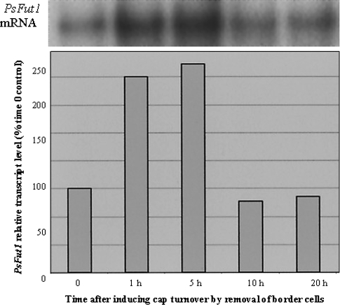
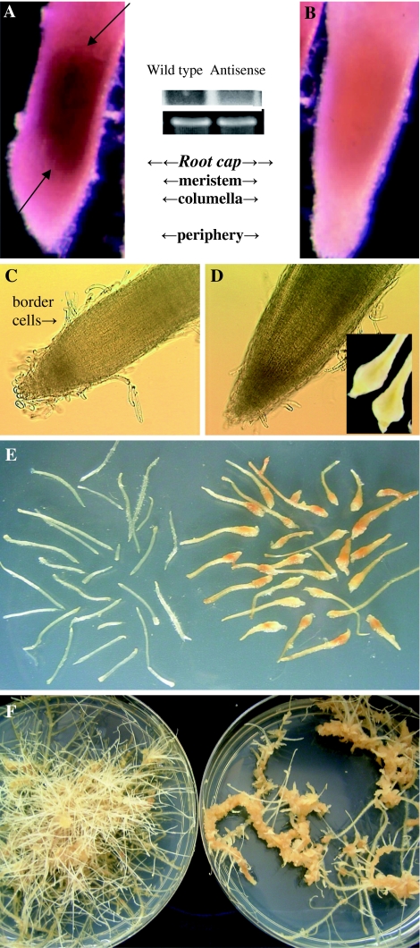
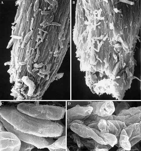
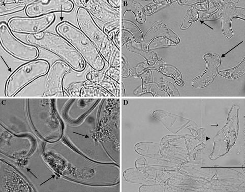
Mitosis and cell wall synthesis in the legume root cap meristem can be induced and synchronized by the nondestructive removal of border cells from the cap periphery. Newly synthesized cells can be examined microscopically as they differentiate progressively during cap development, and ultimately detach as a new population of border cells. This system was used to demonstrate that Pisum sativum L. fucosyl transferase (PsFut1) mRNA expression is strongly expressed in root meristematic tissues, and is induced >2-fold during a 5-h period when mitosis in the root cap meristem is increased. Expression of PsFut1 antisense mRNA in pea hairy roots under the control of the CaMV35S promoter, which exhibits meristem localized expression in pea root caps, resulted in a 50-60% reduction in meristem localized endogenous PsFut1 mRNA expression measured using whole mount in situ hybridization. Changes in gross levels of cell wall fucosylated xyloglucan were not detected, but altered surface localization patterns were detected using whole mount immunolocalization with CCRC-M1, an antibody that recognizes fucosylated xyloglucan. Emerging hairy roots expressing antisense PsFut1 mRNA appeared normal macroscopically but scanning electron microscopy of tissues with altered CCRC-M1 localization patterns revealed wrinkled, collapsed cell surfaces. As individual border cells separated from the cap periphery, cell death occurred in correlation with extrusion of cellular contents through breaks in the wall.


Figures

Fig. 1
Fig. 1
Genomic Southern analysis of pea sequences related to Psfut1. Pea genomic DNA were digested with EcoRI (R1), EcoRV (R5), BamHI (B1), or HindIII (H3) and probed with 32P-labelled Psfut at 55°C
Fig. 2
Fig. 2
Correlation between increased PsFut1 expression and induction of mitosis, which is optimal for 4–5 h after cap turnover is induced (Brigham et al. 1998). Density quantification of the signals obtained from the Northern blot analysis (top panel) was done using the NIH image software program (bottom panel). Relative level was determined based on the ratio of the density values obtained for the signal of interest compared with that of an actin control signal for the same evaluated RNA, and was normalized based on setting Time 0 as 100%
Fig. 3
Fig. 3
Altered mRNA levels occurring in correlation with altered growth and development in transgenic hairy roots expressing PsFut1 antisense mRNA. a, b Whole mount in situ hybridization (WISH); inset; Northern blot analysis of wild type and antisense hairy roots (top), with ribosomal RNA shown to illustrate equal loading (bottom). aPsFut1 expression in wild type hairy roots was localized predominantly within meristematic tissues; antisense mRNA probe (redpurple colour) strongly hybridized to the root cap and apical meristems; b Hybridization was significantly reduced in hairy roots expressing PsFut1 antisense mRNA; c Root tips of wild type hairy roots; d Root tips of hairy roots expressing PsFut1 antisense mRNA. Upon emergence, antisense root tips appear normal. Inset: as roots elongate, the region behind the tip swells; e Appearance of clonal wild type hairy roots (left) or hairy roots expressing Psfut1 antisense mRNA; f Wild type hairy roots grow in culture indefinitely (left); in contrast, antisense hairy roots turn yellow, then brown as they transform into undifferentiated callus (right)
Fig. 4
Fig. 4
Xyloglucan composition (relative amounts) of wild type hairy root tips (R1000), antisense root tips (FUT1), wild type border cells (R1000BC), border cells from a new (<1 week) culture of antisense hairy roots (Fut1NBC), and border cells from an older (>3 weeks) culture of antisense hairy roots (Fut1OldBC) as analyzed by OLIMP (n = 3). The various xyloglucan oligosaccharides (XXG, XXXG, etc.) are presented in their one-letter code (Fry et al. 1993b) including mono- or di-O-acetylated species (+OAc, +2OAc)
Fig. 5
Fig. 5
Aberrant localization of surface fucosylated xyloglucan using CCRC-M1 in root tips of hairy roots expressing PsFut1 antisense mRNA. a Control hairy root expressing PsFut1 antisense mRNA, without exposure to CCRC-M1; a yellow autofluorescence is evident. b Root tip from wild type pea hairy root; strong labelling with CCRC-M1 occurred uniformly over the entire root cap. The bright green fluorescence represents fluorescein-based immunolabelling. c Root tip from individual hairy roots expressing PsFut1 antisense mRNA; bright green fluorescence indicating a positive reaction was evident but varied in intensity among and within individual roots, with some regions exhibiting only yellow autofluorescence (arrow)
Fig. 6
Fig. 6
Scanning electron microscopic comparison of root tips (a, b) versus border cells (c, d) from wild type hairy roots (a, c) versus PsFut1 antisense mRNA lines (b, d)
Fig. 7
Fig. 7
Altered structure and function in cell walls of border cells from three independent lines (PsFut1AS1, PsFut1AS2, PsFut1AS3) of hairy roots expressing PsFut1 antisense mRNA. a Border cells of pea hairy roots, like those of normal roots, have rigid cell walls up to 1 μm in diameter, the nucleus is prominent, and viability is >90%; b Border cells of antisense line PsFut1AS2 were extremely thin and fragile; cells from PsFut1AS3 (inset) were twisted and misshapen, with some wall regions having near-normal width (arrow), adjacent to regions with visible holes (triangle); c Border cells from PsFut1AS1 exhibited regions of structural derangement, appearing to swell (arrows); d Cytoplasmic contents were extruded through holes in the walls of PsFut1AS1 border cells

Similar articles

Cited by

References

    1. {'text': '', 'ref_index': 1, 'ids': [{'type': 'PubMed', 'value': '7958328', 'is_inner': True, 'url': 'https://pubmed.ncbi.nlm.nih.gov/7958328/'}]}
    2. Albersheim P, An JH, Freshour G, Fuller MS, Guillen R, Ham KS, Han MG, Huang J, O’Neil M, Whitecombe A, Williams MV, York WS, Darvill A (1994) Structure and function studies of plant cell wall polysaccharides. Biochem Soc Trans 22:374–378 - PubMed
    1. None
    2. Barlow PW (1975) The root cap. In: Torrey JG, Clarkson DT (eds) The development and function of roots. Academic Press, London, pp 21–54
    1. {'text': '', 'ref_index': 1, 'ids': [{'type': 'DOI', 'value': '10.1104/pp.51.1.174', 'is_inner': False, 'url': 'https://doi.org/10.1104/pp.51.1.174'}, {'type': 'PMC', 'value': 'PMC367376', 'is_inner': False, 'url': 'https://pmc.ncbi.nlm.nih.gov/articles/PMC367376/'}, {'type': 'PubMed', 'value': '16658281', 'is_inner': True, 'url': 'https://pubmed.ncbi.nlm.nih.gov/16658281/'}]}
    2. Bauer WD, Talmadge KW, Keegstra K, Albersheim P (1973) Structure of plant cell walls. 2. Hemicellulose of walls of suspension cultured sycamore cells. Plant Physiol 51:174–187 - PMC - PubMed
    1. {'text': '', 'ref_index': 1, 'ids': [{'type': 'DOI', 'value': '10.1104/pp.118.4.1223', 'is_inner': False, 'url': 'https://doi.org/10.1104/pp.118.4.1223'}, {'type': 'PMC', 'value': 'PMC34738', 'is_inner': False, 'url': 'https://pmc.ncbi.nlm.nih.gov/articles/PMC34738/'}, {'type': 'PubMed', 'value': '9847096', 'is_inner': True, 'url': 'https://pubmed.ncbi.nlm.nih.gov/9847096/'}]}
    2. Brigham LA, Woo HH, Wen F, Hawes MC (1998) Meristem-specific suppression of mitosis and a global switch in gene expression in the root cap of pea by endogenous signals. Plant Physiol 118:1223–1231 - PMC - PubMed
    1. {'text': '', 'ref_index': 1, 'ids': [{'type': 'DOI', 'value': '10.1105/tpc.6.12.1703', 'is_inner': False, 'url': 'https://doi.org/10.1105/tpc.6.12.1703'}, {'type': 'PMC', 'value': 'PMC160556', 'is_inner': False, 'url': 'https://pmc.ncbi.nlm.nih.gov/articles/PMC160556/'}, {'type': 'PubMed', 'value': '12244231', 'is_inner': True, 'url': 'https://pubmed.ncbi.nlm.nih.gov/12244231/'}]}
    2. Brisson LF, Tenhaken R, Lamb C (1994) Function of oxidative cross-linking of cell wall structural proteins in plant disease resistance. Plant Cell 6:1703–1712 - PMC - PubMed

Publication types

MeSH terms