. 2008 Mar 18;9(1):27.
doi: 10.1186/1465-9921-9-27.

IL-13 induces a bronchial epithelial phenotype that is profibrotic

Affiliations

IL-13 induces a bronchial epithelial phenotype that is profibrotic

Nikita K Malavia et al. Respir Res. .

Abstract

Background: Inflammatory cytokines (e.g. IL-13) and mechanical perturbations (e.g. scrape injury) to the epithelium release profibrotic factors such as TGF-beta2, which may, in turn, stimulate subepithelial fibrosis in asthma. We hypothesized that prolonged IL-13 exposure creates a plastic epithelial phenotype that is profibrotic through continuous secretion of soluble mediators at levels that stimulate subepithelial fibrosis.

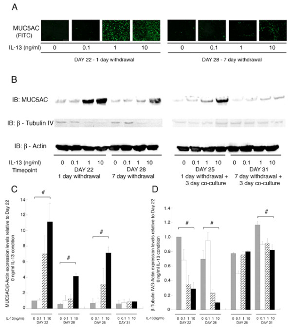
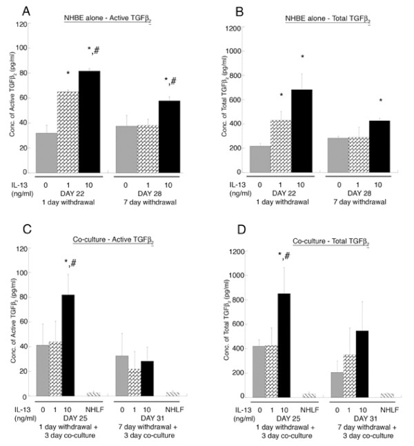
Methods: Normal human bronchial epithelial cells (NHBE) were treated with IL-13 (0, 0.1, 1, or 10 ng/ml) for 14 days (day 7 to day 21 following seeding) at an air-liquid interface during differentiation, and then withdrawn for 1 or 7 days. Pre-treated and untreated NHBE were co-cultured for 3 days with normal human lung fibroblasts (NHLF) embedded in rat-tail collagen gels during days 22-25 or days 28-31.

Results: IL-13 induced increasing levels of MUC5AC protein, and TGF-beta2, while decreasing beta-Tubulin IV at day 22 and 28 in the NHBE. TGF-beta2, soluble collagen in the media, salt soluble collagen in the matrix, and second harmonic generation (SHG) signal from fibrillar collagen in the matrix were elevated in the IL-13 pre-treated NHBE co-cultures at day 25, but not at day 31. A TGF-beta2 neutralizing antibody reversed the increase in collagen content and SHG signal.

Conclusion: Prolonged IL-13 exposure followed by withdrawal creates an epithelial phenotype, which continuously secretes TGF-beta2 at levels that increase collagen secretion and alters the bulk optical properties of an underlying fibroblast-embedded collagen matrix. Extended withdrawal of IL-13 from the epithelium followed by co-culture does not stimulate fibrosis, indicating plasticity of the cultured airway epithelium and an ability to return to a baseline. Hence, IL-13 may contribute to subepithelial fibrosis in asthma by stimulating biologically significant TGF-beta2 secretion from the airway epithelium.


Figures

Figure 1
Figure 1
Protocol for IL-13 treatment and withdrawal. NHBE cells are seeded on Transwells® as described in the Materials and Methods, treatment with IL-13 followed by its withdrawal is carried out as shown. (A) For the control case, the NHBE are cultured in 50:50 epithelial media without any IL-13 all throughout and co-cultured with NHLF embedded in rat tail collagen gel from days 22 to day 25 and day 28 to day 31. (B) NHBE are treated for 14 days from day 7 to day 21 with varying IL-13 concentrations (0.1, 1, 10 ng/ml), then the IL-13 media is withdrawn and replaced with 50:50 epithelial media for 1 day from day 21 to day 22. On day 22 the NHBE are co-cultured with NHLF embedded in a rat-tail collagen gel for a period of 3 days till day 25. (C) The NHBE are treated for 14 days from day 7 to day 21 with varying IL-13 concentrations (0.1, 1, 10 ng/ml), then the IL-13 media is withdrawn and replaced with 50:50 media for a period of 7 days from day 21 to day 28. On day 28 the NHBE are co-cultured with NHLF embedded in a rat-tail collagen gel for 3 days till day 31.
Figure 2
Figure 2
IF staining and Western blot for MUC5AC/β-Tubulin IV in the NHBE. IL-13 mediated a concentration dependent increase in MUC5AC protein levels in the NHBE as seen by (A) Immunofluorescence, where at day 22 and day 28, 1 and 7 days after withdrawal of 14 day treatment with IL-13 (1,10 ng/ml for day 22 and 10 ng/ml for day 28) the staining for MUC5AC is higher compared to the untreated NHBE (0 ng/ml IL-13) (n = 3 donors of NHBE; grown in duplicate; with 3–6 wells per condition; scale bar = 20 μm). DAPI staining of the nuclei showed similar number of cells in all conditions (data not shown). (B) Levels of MUC5AC protein show a dose dependent increase via western blot at day 22 and day 28. Also during co-culture with the NHLF the dose dependent increase of MUC5AC is maintained at day 25 and not at day 31. Levels of β-Tubulin IV protein in the NHBE shown an inverse dependence on IL-13 concentration at days 22 and day 28 with levels remaining constant at day 25 and day 31 of co-culture with NHLF. Images are representative from 3 NHBE donors. (C, D) Quantification of MUC5AC/β-Actin and β-Tubulin IV/β-Actin levels relative to IL-13 concentration of 0 ng/ml at day 22 condition, show a dose dependent increase with IL-13 concentration at day 22,28 and 25 for MUC5AC and dose dependent decrease at day 22, 28 and 31 for β-Tubulin IV (Statistical difference between conditions by ANOVA # p < 0.01).
Figure 3
Figure 3
ELISA for active and total TGF-β2 in the media (A, B). At day 22, the concentration of active and total TGF-β2 in the media of IL-13 pre-treated NHBE at 1 and 10 ng/ml is significantly higher as compared to untreated NHBE (0 ng/ml of IL-13) media; * p < 0.01. At day 22 and day 28, the concentration of active TGF-β2 in the IL-13 pre-treated NHBE at 10 ng/ml is elevated compared to pre-treated NHBE at 1 ng/ml; # p < 0.01. At day 28 active and total TGF-β2 in IL-13 pre-treated NHBE at 10 ng/ml is increased compared to untreated NHBE; * p < 0.01. (C, D) At day 25, the NHBE pre-treated with IL-13 at 10 ng/ml, has higher levels of active and total TGF-β2 in the media as compared to untreated and pre-treated NHBE at 1 ng/ml co-cultured with NHLF (*, # p < 0.01 compared to 0 and 1 ng/ml IL-13 pre-treated NHBE co-cultured with NHLF, respectively). At day 31, there is no significant difference in the levels of active and total TGF-β2 between treatment conditions. NHLF represents levels of active and total TGF-β2 in media of fibroblasts in a collagen gel without NHBE co-culture. All experiments were performed using 3 donors, grown in duplicate, with 3–6 wells per condition.
Figure 4
Figure 4
Quantification of soluble collagen content in the media and matrix. (A) Sircol soluble collagen assay was performed as described in the Materials and Methods, which quantifies the amount of soluble collagen in the cell culture supernatant and newly synthesized salt soluble collagen in the matrix. The amount of soluble collagen secreted in the media at day 25 in the IL-13 pre-treated NHBE at 1 and 10 ng/ml co-cultured with NHLF is augmented as compared to the untreated NHBE co-culture; * p < 0.01 and addition of TGFβ2 neutralizing antibody (10 μg/ml) abolishes this increase (§ p < 0.01 compared to respective condition without TGFβ2 neutralizing antibody). (B) At day 25 there is an increase in newly synthesized salt soluble collagen content in the matrix in the IL-13 pre-treated NHBE at 1 and 10 ng/ml followed by co-culture with NHLF as compared to the untreated NHBE co-culture; * p < 0.01 and the IL-13 pre-treated NHBE at 10 ng/ml co-culture collagen levels are elevated as compared to the IL-13 pretreated NHBE at 1 ng/ml co-culture; # p < 0.01. Also, addition of the TGFβ2 neutralizing antibody abolishes this increase (§ p < 0.01 compared to respective condition without TGFβ2 antibody). The media and matrix collagen levels are normalized to respective levels obtained from NHLF embedded in collagen gels ("NHLF only"). (C, D) Representative Second harmonic generated (SHG) images (scale bar = 50 μm) of collagen fibrils at day 25 are shown along with the quantification of signal intensities. The SHG signals from the collagen secreted by NHLF embedded in rat tail collagen gels which were co-cultured with the IL-13 pre-treated NHBE at 10 ng/ml are elevated compared to the untreated NHBE co-culture; * p < 0.01 and this increase is inhibited on incubation with TGFβ2 neutralizing antibody in the 3 day co-culture period (§ p < 0.01 compared to respective condition without TGFβ2 antibody). Addition of goat IgG did not alter the increased levels of collagen in the matrix and media in the pre-treated NHBE-NHLF co-culture. (E) Exogenous active TGF-β2 at 0.05, 0.1, 0.5, 1 and 10 ng/ml is added in 50:50 epithelial media to NHLF embedded in collagen gels for a period of 3 days. There is a significant increase in the newly synthesized salt soluble collagen content in the matrix with addition of increasing concentration of active TGF-β2 (* p < 0.01 compared to only NHLF condition). All values are normalized to those obtained from "NHLF only" condition. All experiments were performed using 3 donors, grown in duplicate, with 3–6 wells for each condition.

Similar articles

Cited by

References

    1. Cohn L, Elias JA, Chupp GL. Asthma: mechanisms of disease persistence and progression. Annu Rev Immunol. 2004;22:789–815. doi: 10.1146/annurev.immunol.22.012703.104716. - DOI - PubMed
    1. Busse W, Elias J, Sheppard D, Banks-Schlegel S. Airway remodeling and repair. Am J Respir Crit Care Med. 1999;160(3):1035–1042. - PubMed
    1. Davies DE, Wicks J, Powell RM, Puddicombe SM, Holgate ST. Airway remodeling in asthma: new insights. J Allergy Clin Immunol. 2003;111(2):215–25; quiz 226. doi: 10.1067/mai.2003.128. - DOI - PubMed
    1. Elias JA, Zhu Z, Chupp G, Homer RJ. Airway remodeling in asthma. J Clin Invest. 1999;104(8):1001–1006. doi: 10.1172/JCI8124. - DOI - PMC - PubMed
    1. Fixman ED, Stewart A, Martin JG. Basic mechanisms of development of airway structural changes in asthma. Eur Respir J. 2007;29(2):379–389. doi: 10.1183/09031936.00053506. - DOI - PubMed

Publication types

MeSH terms