. 2008 Jul;4(4):783-90.
doi: 10.1016/j.actbio.2008.02.010. Epub 2008 Feb 23.

Non-invasive monitoring of tissue scaffold degradation using ultrasound elasticity imaging

Affiliations

Non-invasive monitoring of tissue scaffold degradation using ultrasound elasticity imaging

Kang Kim et al. Acta Biomater. 2008 Jul.

Abstract

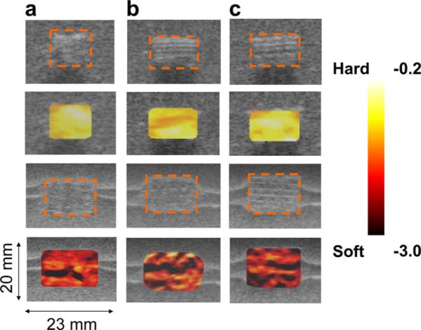
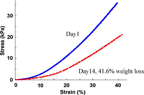
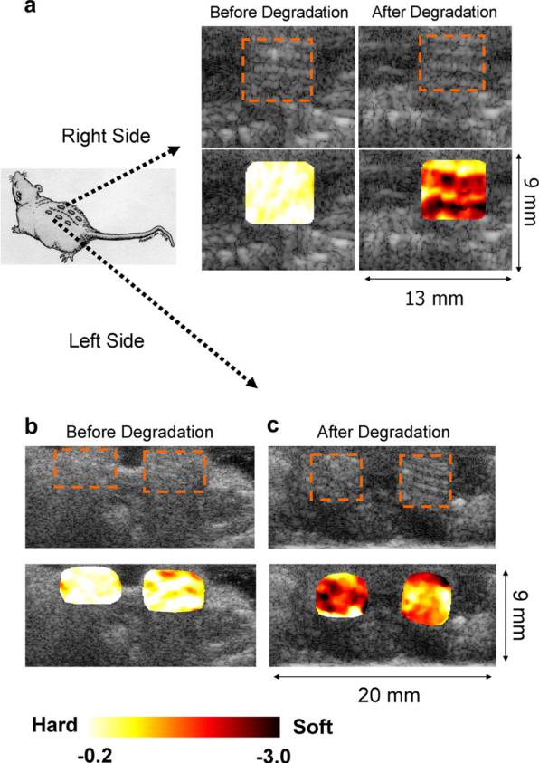
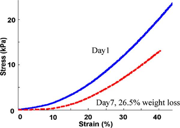
Non-invasively monitoring the extent of cell growth, scaffold degradation and tissue development will greatly help tissue engineers to monitor in vivo regenerate tissue function and scaffold degradation. Currently available methods for tissue and scaffold degradation analysis, such as histology and direct mechanical measurements, are not suitable for continuous monitoring of the same sample in vivo as they destroy cells, tissue matrix and scaffolds. In addition, different samples are prepared and measured at varying times, but high tissue growth deviation between specimens and the need for monitoring tissue growth and scaffold degradation at different times requires large sample numbers for statistical analysis. Ultrasound elasticity imaging (UEI) based on phase-sensitive speckle tracking can characterize the internal structural, compositional and functional change of biomaterial scaffolds and engineered tissues at high resolution. In this study, UEI resolution was 250 microm (axial) by 500 microm (lateral) using a commercial ultrasound transducer centered at 5 MHz. This method allows characterization of both globally and locally altered scaffold and engineered tissue elastic properties. Preliminary in vitro and in vivo results with poly(1,8-octanediol-co-citrate) scaffolds support the feasibility of UEI as a non-invasive quantitative monitoring tool for scaffold degradation and engineered tissue formation. This novel non-invasive monitoring tool will provide direct, time-dependent feedback on scaffold degradation and tissue ingrowth for tissue engineers to improve the design process.


Figures

Fig. 1
Fig. 1
Scaffold fabrication process. HA molds (inverse of the wax molds) are fabricated using thermally curable HA slurry poured into wax molds built on Solidscape machine. The HA molds are then embedded within the pPOC. The pPOC/HA construct inside PTFE mold is post-polymerized at 80 °C for 4 days with vacuum in an oven. The HA + POC construct is removed from the PTFE mold and the HA mold from the construct is dissolved using RDO to obtain the final POC scaffold. The internal structure of the scaffold was examined using micro CT.
Fig. 2
Fig. 2
Experimental set-up. A commercial ultrasound scanner (iU22, Philips) with RF capturing board is used to collect ultrasound frames (a) while the sample (scaffold inside gel phantom or mouse) is deformed by an ultrasound probe connected to a laboratory built deformation device (b).
Fig. 3
Fig. 3
In vitro strain maps overlaid on top of the B-scan for the scaffold samples from the same batch before degradation (first two rows) and after (a) 41.6%, (b) 45.5% and (c) 42.0% degradation by weight (third and fourth rows). The dotted orange boxes represent the scaffolds. The average normalized strain generated inside of the scaffold after the degradation (–2.97 ± 0.74) of about 43% by weight was much higher than before the degradation (–0.43 ± 0.03) of the scaffolds. The coefficient of variation (COV = standard deviation/mean) after the degradation (25%) was much higher than before the degradation (7%). The inhomogeneity of the strain map also reflects the possible heterogeneous distribution of the internal structural degradation. In the lower two columns, the horizontal bright lines around the scaffolds are due to the ultrasound reflection between the layers formed during gel phantom fabrication. These do not affect the mechanical property of the phantom.
Fig. 4
Fig. 4
Direct mechanical measurements for an in vitro sample (in vitro sample 1 in Table 1). The slope of the stress–strain curve for the scaffold before degradation (solid blue) is stiffer than the 41.6% weight degraded scaffold (dashed red). The reconstructed elastic moduli were 89.1 and 38.7 kPa before and after the degradation, respectively. (For interpretation of the references to colour in this figure legend, the reader is referred to the web version of this article.)
Fig. 5
Fig. 5
In vivo strain maps overlaid on top of B-scan for the scaffold samples implanted underneath of the skin on the right (a) and left hand sides (b, c) of the mouse back before and after degradation by weight ((a), 13.6%, left side of (c), 26.5%, and right side of (c) 28.6%). Overall, the scaffolds after the degradation of about 23% by weight generated much higher strains (–1.40 ± 0.24) than those before the degradation (–0.21 ± 0.01). The COV before the degradation (4.8%) was increased to 17% after the degradation. The inhomogeneity of the strain map also reflects the possible heterogeneous distribution of the structural degradation.
Fig. 6
Fig. 6
Direct mechanical measurements for an in vivo sample (in vivo sample 2 in Table 1). The slope of the stress–strain curve for the scaffold before degradation (solid blue) is stiffer than after 26.5% weight degradation (dashed red). The reconstructed elastic moduli are 56.4 and 38.1 kPa before and after the degradation, respectively. (For interpretation of the references to colour in this figure legend, the reader is referred to the web version of this article.)

References

    1. Gao J, Crapo PM, Wang Y. Macroporous elastomeric scaffolds with extensive micropores for soft tissue engineering. Tissue Eng. 2006;12:917–25. - PubMed
    1. Sun W, Darling A, Starly B, Nam J. Computer-aided tissue engineering: overview, scope and challenges. Biotechnol Appl Biochem. 2004;39:29–47. - PubMed
    1. Lerner RM, Parker KJ, Holen J, Gramiak R, Waag RC. Sono-elasticity: medical elasticity images de rived from ultra sound signals in mechanically vibrated targets. Acoust Imaging. 1988;16:317–27.
    1. Ophir J, Céspedes I, Ponnekanti H, Yazdi Y, Li X. Elastography: a quantitative method for imaging the elasticity of biological tissues. Ultrasonic Imaging. 1991;13:111–34. - PubMed
    1. Skovoroda AR, Emelianov SY, Lubinski MA, Sarvazyan AP, O'Donnell M. Theoretical analysis and verification of ultra sound displacement and strain imaging. IEEE Trans UFFC. 1994;41:302–13.

Publication types

LinkOut - more resources