. 2008 May 16;283(20):13762-70.
doi: 10.1074/jbc.M710592200. Epub 2008 Mar 18.

Crystal structure of a functional dimer of the PhoQ sensor domain

Affiliations

Crystal structure of a functional dimer of the PhoQ sensor domain

Jonah Cheung et al. J Biol Chem. .

Abstract

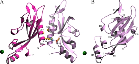
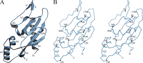
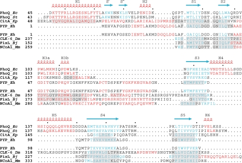
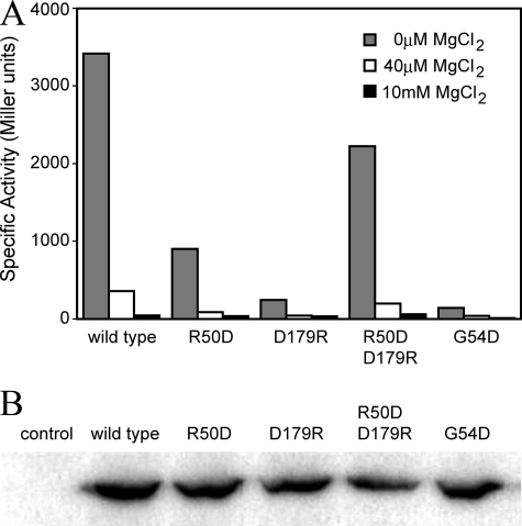
The PhoP-PhoQ two-component system is a well studied bacterial signaling system that regulates virulence and stress response. Catalytic activity of the histidine kinase sensor protein PhoQ is activated by low extracellular concentrations of divalent cations such as Mg2+, and subsequently the response regulator PhoP is activated in turn through a classic phosphotransfer pathway that is typical in such systems. The PhoQ sensor domains of enteric bacteria contain an acidic cluster of residues (EDDDDAE) that has been implicated in direct binding to divalent cations. We have determined crystal structures of the wild-type Escherichia coli PhoQ periplasmic sensor domain and of a mutant variant in which the acidic cluster was neutralized to conservative uncharged residues (QNNNNAQ). The PhoQ domain structure is similar to that of DcuS and CitA sensor domains, and this PhoQ-DcuS-CitA (PDC) sensor fold is seen to be distinct from the superficially similar PAS domain fold. Analysis of the wild-type structure reveals a dimer that allows for the formation of a salt bridge across the dimer interface between Arg-50' and Asp-179 and with nickel ions bound to aspartate residues in the acidic cluster. The physiological importance of the salt bridge to in vivo PhoQ function has been confirmed by mutagenesis. The mutant structure has an alternative, non-physiological dimeric association.


Figures

FIGURE 1.
FIGURE 1.
Structure of the wild-type PhoQ 43–190 dimer in complex with nickel. A, the overall structure in complex with nickel is shown as a ribbon diagram. Nickel ions are shown as green spheres. The Arg-50′ → Asp-179 salt bridge is shown with residue side chains depicted in a stick representation (carbon colored yellow, nitrogen colored blue, and oxygen colored red). A line of black dots marks interacting atoms of the salt bridge. The Cα atoms of Gly-54 residues are shown as yellow spheres. Residues are identified by one-letter-code labels. Colored dots are drawn to represent a hypothetical path of the protein backbone through disordered regions corresponding to residues 135 and 136 of molecule A (light purple), and residues 136 and 137 of molecule B (light red). B, the structure of one subunit (molecule A) of the wild-type PhoQ 43–190 dimer is shown from a 90° rotation about the vertical axis of the dimer as shown in A. Secondary structure elements are labeled in black. The diagram was created using MolScript (43).
FIGURE 2.
FIGURE 2.
Structure of an acid mutant PhoQ 43–190 subunit. A, the overall structure of the acid mutant PhoQ 43–190 (molecule A) is shown as a ribbon diagram with secondary structure elements labeled in black. B, a stereo plot of the Cα trace of the same acid mutant PhoQ 43–190 molecule. Every tenth Cα atom is depicted as a black sphere and labeled accordingly. The diagrams were created using MolScript (43) and BobScript (44).
FIGURE 3.
FIGURE 3.
Structure-based sequence alignments. Structure-based sequence alignments comparing PDC to PAS domains. Residues in helices are colored red, and residues in strands are colored blue. Secondary structure elements of E. coli PhoQ are also shown and labeled above the alignments, with helices in red and strands in blue.Inthe top set, the PDC sensor domains PhoQ and CitA are aligned with the PYP PAS domain. Regions in CitA and PYP having Cα positions structurally aligned with those in E. coli PhoQ are shaded in light gray.Inthe bottom set, the PAS domains CLK-6, FixL, and NcoA-1 are aligned with PYP. Regions in CLK-6, FixL, and NcoA-1 having Cα positions structurally aligned with those of PYP are shaded in light gray. Organism names are abbreviated in italics (Ec for E. coli, St for S. typhimurium, Kp for Klebsiella pneumoniae, Eh for Ectothiorhodospira halophila, Dm for Drosophila melanogaster, Bj for Bradyrhizobium japonicum, and Mm for Mus musculus).
FIGURE 4.
FIGURE 4.
In vivo activity and relative expression levels of PhoQ. A, E. coli strain CSH26ΔQ/FlacIQkan, containing the pNL3 reporter plasmid and pLPQ2 for expressing the wild-type and mutant PhoQ proteins, were assayed for β-galactosidase activity following growth in N-media (0 μm MgCl2) or as supplemented with 40 μm or 10 mm MgCl2. Specific activity is shown in Miller units. Standard deviations are <10% of the mean, calculated from at least three individual experiments. B, the relative expression levels of the wild-type and mutant PhoQ proteins are shown by Western blotting, probed using an antibody specific for the cytoplasmic domain of PhoQ. The PhoQ variants are labeled above each lane accordingly. The control is the E. coli strain containing the “empty” pLPQ2 vector lacking the phoP-phoQ operon.

References

    1. Stock, A. M., Robinson, V. L., and Goudreau, P. N. (2000) Annu. Rev. Biochem. 69 183-215 - PubMed
    1. West, A. H., and Stock, A. M. (2001) Trends Biochem. Sci. 26 369-376 - PubMed
    1. Wolanin, P. M., Thomason, P. A., and Stock, J. B. (2002) Genome Biology http://genomebiology.com/2002/3/10/reviews/3013 - PMC - PubMed
    1. Robinson, V. L., Buckler, D. R., and Stock, A. M. (2000) Nat. Struct. Biol. 7 626-633 - PubMed
    1. Dutta, R., Qin, L., and Inouye, M. (1999) Mol. Microbiol. 34 633-640 - PubMed

Publication types

LinkOut - more resources