. 2008 May 23;283(21):14581-9.
doi: 10.1074/jbc.M707733200. Epub 2008 Mar 18.

Hepatocyte-specific c-Met deletion disrupts redox homeostasis and sensitizes to Fas-mediated apoptosis

Affiliations

Hepatocyte-specific c-Met deletion disrupts redox homeostasis and sensitizes to Fas-mediated apoptosis

Luis E Gómez-Quiroz et al. J Biol Chem. .

Abstract

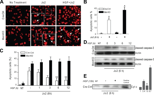
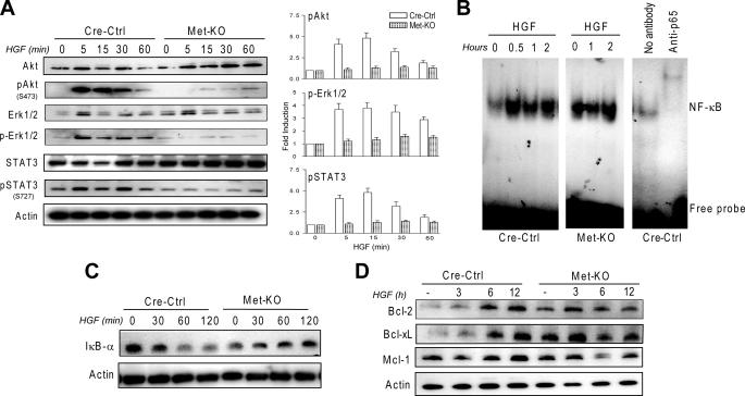
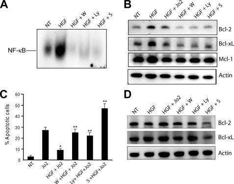
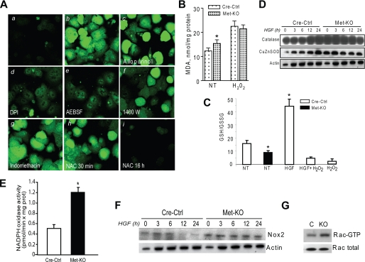
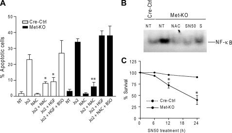
The hepatocyte growth factor and its receptor c-Met direct a pleiotropic signal transduction pathway that controls cell survival. We previously demonstrated that mice lacking c-Met (Met-KO) in hepatocytes were hypersensitive to Fas-induced liver injury. In this study, we used primary hepatocytes isolated from Met-KO and control (Cre-Ctrl) mice to address more directly the protective effects of c-Met signaling. Loss of c-Met function increased sensitivity to Fas-mediated apoptosis. Hepatocyte growth factor suppressed apoptosis in Cre-Ctrl but not Met-KO hepatocytes concurrently with up-regulation of NF-kappaB and major antiapoptotic proteins Bcl-2 and Bcl-xL. Intriguingly, Met-KO hepatocytes exhibited intrinsic activation of NF-kappaBas well as Bcl-2 and Bcl-xL. Furthermore, unchallenged Met-KO cells displayed oxidative stress as evidenced by overproduction of reactive oxygen species, which was associated with greater NADPH and Rac1 activities, was blocked by the known NADPH oxidase inhibitors, and was paralleled by increased lipid peroxidation and reduced glutathione (GSH) content. N-Acetylcysteine, an antioxidant and GSH precursor, significantly reduced Jo2-induced cell death. Conversely, the GSH-depleting agent buthionine sulfoximine completely abolished the protective effects of N-acetylcysteine in Met-KO hepatocytes. In conclusion, genetic inactivation of c-Met in mouse hepatocytes caused defects in redox regulation, which may account for the increased sensitivity to Fas-induced apoptosis and adaptive up-regulation of NF-kappaB survival signaling. These data provide evidence that intact c-Met signaling is a critical factor in the protection against excessive generation of endogenous reactive oxygen species.


Figures

FIGURE 1.
FIGURE 1.
Lack of c-Met function increases sensitivity of mouse hepatocytes to Jo2-mediated apoptosis. A, representative images of primary hepatocytes stained with PI. Cre-Ctrl and Met-KO cultures were serum-starved for 16 h and incubated with Jo2 (0.5 μg/ml) for 6 h in the absence or presence of HGF (40 ng/ml) for 12 h. Insets show nuclear condensation, DNA fragmentation, and apoptotic body formation characteristic of apoptosis (white arrowheads). Original magnification, ×200. B, apoptotic index in Cre-Ctrl and Met-KO hepatocytes treated with Jo2 (0.5 μg/ml) for 6 h after serum starvation for 16 h. Each column represents the mean ± S.E. At least 500 nuclei were counted from duplicate cultures in three independent experiments. *, p < 0.05 against respective untreated cultures. C, time course of HGF protection against Jo2-induced apoptosis. Serum-starved (16 h) Cre-Ctrl and Met-KO hepatocytes were incubated with Jo2 (0.5 μg/ml) in the absence or presence of HGF (40 ng/ml) for the indicated time. Apoptotic index was detected by counting apoptotic cells after PI staining. Each column represents the mean ± S.E. At least 500 nuclei were counted from duplicate cultures in three independent experiments. *, p < 0.05 against respective Met-KO culture. **, p < 0.05 against Cre-Ctrl in the absence of HGF pretreatment. NT, no treatment. D, activation of caspase 3. Whole cell lysates from Cre-Ctrl and Met-KO hepatocytes were immunoblotted with anti-cleaved caspase-3. Treatment with HGF and Jo2 was performed as described in C. E, cytochrome c release. Cre-Ctrl hepatocytes were serum-starved for 16 h and incubated with Jo2 (0.5 μg/ml) for 6 h in the absence or presence of HGF (40 ng/ml) for 12 h. Cytosol fractions were immunoblotted with anti-cytochrome c (Cyt c). Mitochondrial protein was used as positive control. The intensity of each band was quantified by densitometry and expressed as fold of control (NT). A representative Western blot of three experiments is shown in D and E.*, p < 0.05 versus treated with Jo2.
FIGURE 2.
FIGURE 2.
Met-KO primary hepatocytes exhibit constitutive activation of NF-κB and antiapoptotic proteins. A, HGF activates Akt, Erk1/2, and STAT3 in Cre-Ctrl but not Met-KO cells. Whole cell lysates prepared from serum-starved (16 h) Cre-Ctrl and Met-KO cultures treated with 40 ng/ml HGF for 0, 5, 15, 30, and 60 min were immunoblotted with anti-AKT, anti-Erk1/2, anti-Stat3, and phospho-specific antibodies against Akt Ser(P)-473, Erk1/2 Thr(P)-202/Tyr(P)-204, and Stat3 Ser(P)-727. The intensity of each protein was quantified by densitometry normalized to actin. The density value of total protein in untreated samples was designated as 1. Each bar represents the mean ± S.E. B, electromobility shift assay of NF-κB DNA binding activity in serum-starved (16 h) Cre-Ctrl and Met-KO hepatocytes treated with HGF (40 ng/ml) for the indicated time. The presence of supershift complex with anti-p65 demonstrates the assay specificity. C, kinetics of IκB degradation. Whole cell lysates prepared from serum-starved (16 h) Cre-Ctrl and Met-KO hepatocytes treated with 40 ng/ml HGF for 0, 30, 60, and 120 min were immunoblotted with anti-IκB-α. D, expression of antiapoptotic proteins in Cre-Ctrl and Met-KO cultures. Cre-Ctrl and Met-KO hepatocytes were serum-starved for 16 h and incubated with HGF (40 ng/ml) for the indicated time. Whole cell lysates were immunoblotted with anti-Bcl-2, anti-Bcl-xL, and anti-Mcl-1. A representative Western blot of three experiments is shown in A, C, and D.
FIGURE 3.
FIGURE 3.
NF-κB activation is required for the antiapoptotic effects of HGF in Cre-Ctrl hepatocytes. A, electromobility shift assays of NF-κB DNA binding activity. Cre-Ctrl hepatocytes were pretreated with 100 μm wortmannin (W), 50 μm LY294002 (Ly), and 100 μm sulfasalazine (S) for 30 min followed by 40 ng/ml HGF for 12 h. Results were confirmed in three independent experiments. B, expression of antiapoptotic proteins in Cre-Ctrl cells. Cells were pretreated with 100 μm wortmannin, 50 μm LY294002, and 100 μm sulfasalazine for 30 min followed by 40 ng/ml HGF for 12 h. C, effect of inhibition of PI3K/Akt and NF-κB on frequency of apoptosis. After 16 h of serum starvation, cells were pretreated with 100 μm wortmannin, 50 μm LY294002, or 100 μm sulfasalazine for 30 min followed by treatment with HGF (40 ng/ml) for 12 h and Jo2 (0.5 μg/ml) for the last 6 h in culture. Apoptotic index was detected by counting apoptotic cells after PI staining. Each column represents the mean ± S.E. At least 500 nuclei were counted from duplicate slides from three independent cultures. D, expression of antiapoptotic proteins in Met-KO cells. Cells were pretreated with 100 μm wortmannin, 50 μm LY294002, and 100 μm sulfasalazine for 30 min followed by 40 ng/ml HGF for 12 h. A representative Western blot of three experiments is shown in B and D.*, p < 0.05 against Jo2 treatment alone; **, p < 0.05 against HGF + Jo2 treatment. NT, no treatment.
FIGURE 4.
FIGURE 4.
Met-KO hepatocytes are subjected to oxidative stress. A, Cre-Ctrl (panel a) and Met-KO (panel b) cells were incubated with the oxidative-sensitive probe DCFH-AM (3 μm) for 30 min. Met-KO cells were pretreated with 100 μm allopurinol (panel c), 100 μm DPI (panel d), 250 μm 4-(2-aminoethyl) benzenesulfonyl fluoride (AEBSF) (panel e), 100 μm N-(3-(aminomethyl)benzyl) acetamidine (1400W) (panel f), 100 μm indomethacin (panel g), and 10 mm NAC (panel h) for 30 min and 16 h (panel i) before incubation with 3 μm DCFH-AM for 30 min. Original magnification, ×100. B, lipid peroxidation was assessed by malondialdehyde (MDA) content in Cre-Ctrl and Met-KO hepatocytes after serum starvation for 16 h. Treatment with 0.25 mm H2O2 was used as positive control. Each column represents the mean ± S.E. of three independent experiments. *, p < 0.05 against Cre-Ctrl. C, ratio of reduced glutathione (GSH) to oxidized glutathione (GSSG) determined by HPLC in serum-starved Cre-Ctrl and Met-KO hepatocytes. Cre-Ctrl hepatocytes were treated with H2O2 (0.25 mm) for 6 h as positive control. Each column represents the mean ± S.E. of three independent experiments. *, p < 0.05 against untreated Cre-Ctrl. NT, no treatment. D, expression of antioxidant proteins. Whole cell lysates prepared from Cre-Ctrl and Met-KO hepatocytes treated with HGF (40 ng/ml) at 0, 3, 6, 12, and 24 h were immunoblotted with anti-catalase and anti-CuZn superoxide dismutase. A representative Western blot of three experiments is shown. E, NADPH oxidase activity. F, expression of Nox2. Whole cell lysates prepared and treated as described above (D) were immunoblotted with anti-Nox2. G, Rac1 activation assay. PAK-1 was used to pull down the activated Rac-GTP from Cre-Ctrl (C) and Met-KO (KO) cell lysates. Immunoblotting was performed using Rac1 monoclonal antibody to reveal the amount of total Rac1.
FIGURE 5.
FIGURE 5.
Reduced GSH levels in Met-KO hepatocytes increase sensitivity to apoptosis. A, Cre-Ctrl and Met-KO hepatocytes were pretreated with BSO (300 μm) for 1 h before incubation with HGF (40 ng/ml) or NAC (10 mm) for 12 h followed by Jo2 (0.5 μg/ml) treatment for 6 h. Apoptotic index was determined by counting apoptotic cells after propidium iodine staining. Each column represents the mean ± S.E. At least 500 nuclei were counted from duplicate cultures from three independent experiments. *, p < 0.05 against Cre-Ctrl cells treated with Jo2 alone or Jo2 + HGF + BSO; **, p < 0.05 against Met-KO treated with Jo2 alone, Jo2 + HGF, or Jo2 + NAC + BSO. B, electromobility shift assays of NF-κB DNA binding activity was performed using nuclear extracts from untreated (NT) Cre-Ctrl cells and Met-KO cells and Met-KO cells treated with 10 mm NAC, 30 μm SN50, or 100 μm sulfasalazine (S) for 12 h. C, cell viability in Cre-Ctrl and Met-KO hepatocytes treated with 30 μm SN50 for 24 h as determined by crystal violet assay. Mean ± S.E. of three independent experiments are shown. *, p < 0.05 against respective Cre-Ctrl cells.

Similar articles

Cited by

References

    1. Peter, M. E., and Krammer, P. H. (2003) Cell Death Differ. 10 26-35 - PubMed
    1. Ogasawara, J., Watanabe-Fukunaga, R., Adachi, M., Matsuzawa, A., Kasugai, T., Kitamura, Y., Itoh, N., Suda, T., and Nagata, S. (1993) Nature 364 806-809 - PubMed
    1. Gao, C. F., and Vande Woude, G. F. (2005) Cell Res. 15 49-51 - PubMed
    1. Comoglio, P. M. (2001) Nat. Cell Biol. 3 E161-E162 - PubMed
    1. Matsumoto, K., and Nakamura, T. (1996) J. Biochem. (Tokyo) 119 591-600 - PubMed

Publication types

MeSH terms