. 2008 May 16;283(20):14100-8.
doi: 10.1074/jbc.M801357200. Epub 2008 Mar 17.

Differential regulation of the IL-17 receptor by gammac cytokines: inhibitory signaling by the phosphatidylinositol 3-kinase pathway

Affiliations

Differential regulation of the IL-17 receptor by gammac cytokines: inhibitory signaling by the phosphatidylinositol 3-kinase pathway

Matthew J Lindemann et al. J Biol Chem. .

Abstract

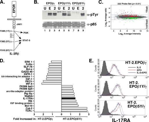
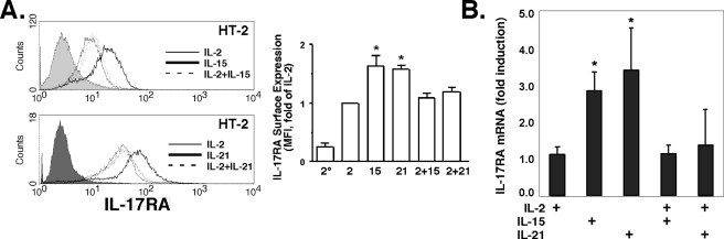
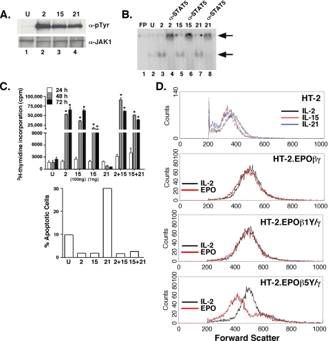
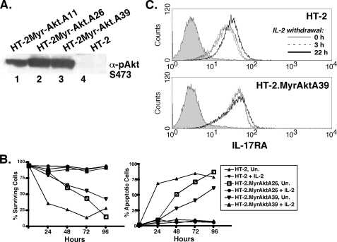
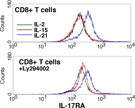
The gammac-family cytokine IL-2 activates signaling events that contribute to cell survival and proliferation, the best-studied of which are the STAT-5 and phosphatidylinositol 3-kinase (PI3K) pathways. The starting point of this study was to define genes regulated by the IL-2R-mediated PI3K pathway in T cells. Accordingly, we used an erythropoietin (EPO) receptor chimeric receptor system in which IL-2-dependent HT-2 T cells expressed a mutant EPO-IL-2Rbeta construct where Tyr-338 is mutated to Phe. Cells expressing this mutant IL-2Rbeta chain fail to induce phosphorylation of PI3K-p85alpha/beta or activate Akt, but mediate normal IL-2-dependent proliferation and activation of JAK1 and STAT-5A/B. Microarray analyses revealed differential regulation of numerous genes compared with cells expressing a wild-type IL-2Rbeta, including up-regulation of the IL-17 receptor subunit IL-17RA. Blockade of the PI3K pathway but not p70S6K led to up-regulation of IL-17RA, and constitutive Akt activation was associated with suppressed IL-17RA expression. Moreover, similar to the mutant EPO-IL-2Rbeta chimera, IL-15 and IL-21 induced IL-17RA preferentially compared with IL-2, and IL-2 but not IL-15 or IL-21 mediated prolonged activation of the PI3K p85 regulatory subunit. Thus, there are intrinsic signaling differences between IL-2 and IL-15 that can be attributed to differences in activation of the PI3K pathway.


Figures

FIGURE 1.
FIGURE 1.
Differential gene expression mediated by the PI3K pathway through IL-2RβY338. A, schematic diagram of the EPOR-IL-2Rβ chimeric receptor construct. The location of JAK-binding site (Box1/Box2) and the pathways activated by individual tyrosine residues are indicated. B, p85 subunit of PI3K is activated through Tyr-338 (1Y). HT-2 cells expressing the indicated EPO-IL-2R chimeras were stimulated for 1 h with EPO or IL-2. Lysates were immunoprecipitated with α-p85α/β Abs, separated by SDS-PAGE, and immunoblotted with α-Ptyr (4G10) Abs (top) or p85α/β Abs (bottom). C, differentially expressed genes in HT-2.EPOβγ cells versus HT-2.EPOβ5Y/γ cells. Gene differences with a p value <0.01 are shown (222 total genes). D, selected genes enhanced in HT-2.EPOβ5Y/γ cells or HT-2.EPOβγ cells. E, IL-17RA surface expression is elevated in HT-2.EPOβ5Y/γ cells. The indicated cell lines were incubated in EPO for 24 h and IL-17RA expression was assessed by flow cytometry. Filled histograms, isotype control.
FIGURE 2.
FIGURE 2.
IL-17RA is induced by IL-15 and IL-21 but not by IL-2. A, IL-2 dominantly suppresses IL-17RA surface expression mediated by IL-15 and IL-21. HT-2 cells were treated for 24 h with the indicated cytokines, and IL-17RA surface expression was assessed by flow cytometry. Filled histograms, isotype control. Average mean fluorescent intensity of IL-17RA expression relative to IL-2-treated cells was determined from five independent experiments (right panel). , secondary Ab alone. B, IL-2 dominantly suppresses IL-17RA mRNA expression mediated by IL-15 and IL-21. HT-2 cells were treated for 24 h with the indicated cytokines, and IL-17RA mRNA levels were determined in triplicate by real-time RT-PCR. *, p < 0.05 compared with IL-2-treated control.
FIGURE 3.
FIGURE 3.
IL-2 and IL-15 differentially activate the PI3K pathway but not the JAK-STAT-5 pathway or proliferation in HT-2 cells. A, IL-2, IL-15, and IL-21 activate JAK1 equivalently in HT-2 cells. HT-2 cells were stimulated for 15 min with the indicated cytokines. Lysates were immunoprecipitated with α-JAK1 Abs, separated by SDS-PAGE, and immunoblotted with α-PTyr (4G10) Abs (top) or α-JAK1 Abs (bottom). B, IL-2, IL-15 and IL-21 activate STAT-5 equivalently in HT-2 cells. HT-2 cells were stimulated for 15 min with the indicated cytokines. Nuclear extracts were subjected to EMSA with a 32P-labeled STAT oligonucleotide (7) (which binds to STAT-1, 5, 6 and to a lesser extent STAT-3). FP, free probe. Supershifting with α-STAT-5 Abs was performed in lanes 4, 6, and 8. Arrows indicate shifted and supershifted complexes. C, IL-2, IL-15, but not IL-21 drive proliferation and inhibit apoptosis in HT-2 cells. HT-2 cells were cultured for 24, 48, or 72 h in the indicated cytokines and concentrations. Proliferation was assessed by [3H]thymidine incorporation in triplicate ± S.D., *, p < 0.05. Apoptosis was determined by FACS staining with propidium iodide and GFP-Annexin V, and apoptotic cells were defined as PI-negative/GFP-Annexin V-positive, as described in Ref. , . Data are representative of three experiments. D, signaling from PI3K via IL-2 and Y338 promotes increased cell size. HT-2 cells (top panel) or HT-2 cells expressing the indicated EPO-IL-2Rβ chimeras (bottom 3 panels) were cultured in the indicated cytokines for 24 h, and cell size was assessed by forward scatter analysis by flow cytometry.
FIGURE 4.
FIGURE 4.
The PI3K pathway inhibits IL-17RA expression. A, sustained phosphorylation of p85 induced by IL-2 but not IL-15 or IL-21. HT-2 cells were cultured in IL-2, IL-15, or IL-21 for the indicated time points, and phosphorylation of the p85α/β was assessed as in Fig. 1. B, HT-2 cells express PTEN and SHIP. Lysates from HT-2, Jurkat T cells or the A20 B cell line were immunoblotted with Abs to PTEN or IKKγ (top panels) or immunoprecipitated with α-SHIP Abs and probed for SHIP (bottom panel). C, PI3K inhibitors up-regulate IL-17RA. HT-2 cells were incubated in IL-2 ± LY294002 or Wortmannin for 20 h. IL-17RA expression was assessed by flow cytometry. D, rapamycin inhibits p70S6K but not IL-15-induced IL-17RA expression. HT-2 cells were incubated with IL-2 or IL-15 ± Rapamycin for 20 h. IL-17RA (top) or p70S6K (bottom) were assessed by flow cytometry.
FIGURE 5.
FIGURE 5.
A constitutively active form of Akt promotes IL-17RA expression. A, HT-2 cells stably express Myr-Akt. Lysates from 3 HT-2 subcloned stably transfected with Myr-Akt were immunoblotted with α-pAkt (Ser-473). B, Myr-Akt delays apoptosis induced by cytokine withdrawal. HT-2 cells expressing Myr-Akt (clones A26 and A39) were incubated with or without IL-2 for the indicated time points, and cell survival (PI-negative) or apoptosis (GFP-Annexin V-positive) cells were assessed by flow cytometry. C, expression of a constitutively active form of Akt prevents up-regulation of IL-17RA induced by cytokine withdrawal. HT-2 or HT-2.MyrAktA39 cells were deprived of IL-2 for the indicated times, and IL-17RA expression was assessed by flow cytometry.
FIGURE 6.
FIGURE 6.
Regulation of IL-17RA in primary CD8+ T cells. Splenic CD8+ T cells were purified by magnetic beads and stimulated for 6 h with the indicated cytokines with DMSO (top) or LY294002 (bottom). IL-17RA was assessed by flow cytometry.
FIGURE 7.
FIGURE 7.
IL-17 does not induce detectable signaling in HT-2 cells or primary T lymphocytes. A and B, IL-17 does not induce canonical target genes such as IL-6 or LIX/CXCL5. HT-2 cells, splenocytes, or purified CD8+ T cells were incubated with the indicated cytokines for 2-24 h, and conditioned supernatants were assayed in triplicate for IL-6 and LIX/CXCL5 by ELISA (36).

Similar articles

Cited by

References

    1. Ozaki, K., and Leonard, W. J. (2002) J. Biol. Chem. 277 29355-29358 - PubMed
    1. Ma, A. (2000) Mod. Asp. Immunobiol. 1 102-104
    1. Waldmann, T. (2006) Nat. Rev. 6 595-601 - PubMed
    1. Johnston, J. A., Kawamura, M., Kirken, R. A., Chen, Y. Q., Blake, T. B., Shibuya, K., Ortaldo, J. R., McVicar, D. W., and O'Shea, J. J. (1994) Nature 370 151-153 - PubMed
    1. Russell, S. M., Johnston, J. A., Noguchi, M., Kawamura, M., Bacon, C. M., Friedmann, M., Berg, M., McVicar, D. W., Whitthuhn, B. A., Silvennoinen, O., Goldman, A. S., Schmalsteig, F. C., Ihle, J. N., O'Shea, J. J., and Leonard, W. J. (1994) Science 266 1042-1045 - PubMed

Publication types

LinkOut - more resources