. 2008 May 30;283(22):15381-9.
doi: 10.1074/jbc.M710296200. Epub 2008 Mar 17.

E2F1 regulates the base excision repair gene XRCC1 and promotes DNA repair

Affiliations

E2F1 regulates the base excision repair gene XRCC1 and promotes DNA repair

Dexi Chen et al. J Biol Chem. .

Abstract

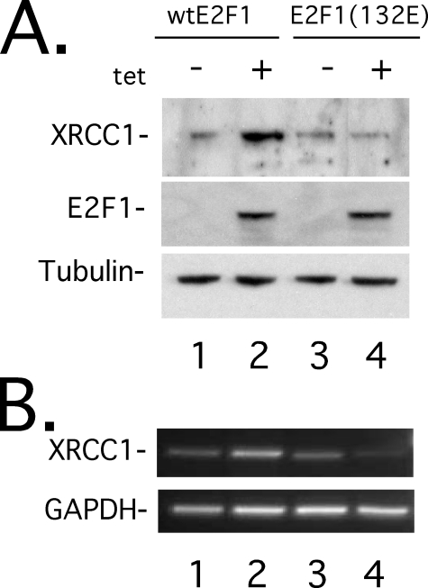
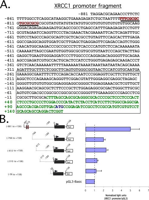
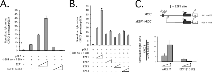
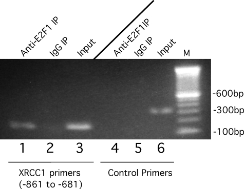
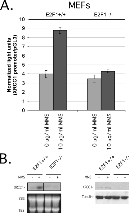
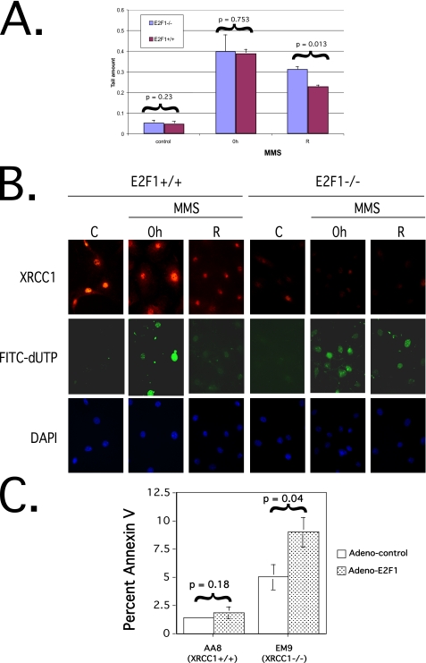
The E2F1 transcription factor activates S-phase-promoting genes, mediates apoptosis, and stimulates DNA repair through incompletely understood mechanisms. XRCC1 (x-ray repair cross-complementing group 1) protein is important for efficient single strand break/base excision repair. Although both damage and proliferative signals increase XRCC1 levels, the mechanisms regulating XRCC1 transcription remain unclear. To study these upstream mechanisms, the XRCC1 promoter was cloned into a luciferase reporter. Ectopic expression of wild-type E2F1, but not an inactive mutant E2F1(132E), activated the XRCC1 promoter-luciferase reporter, and deletion of predicted E2F1 binding sites in the promoter attenuated E2F1-induced activation. Endogenous XRCC1 expression increased in cells conditionally expressing wild-type, but not mutant E2F1, and methyl methanesulfonate-induced DNA damage stimulated XRCC1 expression in E2F1(+/+) but not E2F1(-/-) mouse embryo fibroblasts (MEFs). Additionally, E2F1(-/-) MEFs displayed attenuated DNA repair after methyl methanesulfonate-induced damage compared with E2F1(+/+) MEFs. Moreover, Chinese hamster ovary cells with mutant XRCC1 (EM9) were more sensitive to E2F1-induced apoptosis compared with Chinese hamster ovary cells with wild-type XRCC1 (AA8). These results provide new mechanistic insight into the role of the E2F pathway in maintaining genomic stability.


Figures

FIGURE 1.
FIGURE 1.
E2F1 expression stimulates endogenous XRCC1. Shown are Western blots (A) and ethidium bromide-stained agarose gel (B) of semiquantitative reverse transcription-PCR, on Saos2 cell lines conditionally expressing wild-type E2F1 (lanes 1 and 2) or DNA binding-incompetent mutant E2F1(132E) (lanes 3 and 4). Prior to induction, cell lines were maintained in 0.5% fetal bovine serum-containing medium for 30 h, induced with 2.0 μg/ml doxycycline, and then harvested 24 h later.
FIGURE 2.
FIGURE 2.
XRCC1 promoter fragment sequence and luciferase reporter assay define an active promoter region. A, genomic sequence from -881 to +158 relative to the predicted transcription start site/cDNA 5′-end indicated as +1. Exon 1 is indicated in green type, and the translation-initiation codon is shown in blue type. The open box indicates a CpG island. Underlined sequences in red denote putative E2F-binding sites. B (left), schematic representation of a series of promoter deletion mutants in pGL3-Basic-luciferase reporter. The red boxes denote putative E2F-binding sites. The black box represents exon 1. An open box with [luc] denotes luciferase reporter. Right, luciferase readout of the indicated promoter-reporters after transfection into Saos2 cells. Light units normalized to a β-galactosidase signal. In all, 1.0 μg of the indicated promoter-reporter and pRSV β-galactosidase plasmids was used for all transfections. S.D. of triplicate experiments is shown.
FIGURE 3.
FIGURE 3.
E2F stimulates the XRCC1 promoter-luciferase reporter. A, relative light units, normalized to a β-galactosidase signal, of pGL3-XRCC1-(-881 to +158) (0.1 μg) co-transfected with increasing amounts of wild-type E2F1 or mutant E2F1(132E) expression plasmids (50-250 ng) into Saos2 cells. B, relative light units, normalized to a β-galactosidase signal, of pGL3-XRCC1-(-881 to +158) (0.1 μg) co-transfected with increasing amounts of the indicated E2F expression vectors (50-250 ng) into Saos2 cells. C, relative light units, normalized to a β-galactosidase signal, of the relative -fold change of the XRCC1-(-881 to +158) promoter-reporter versus an E2F binding site-deleted XRCC1 promoter-reporter (ΔE2F1-XRCC1), in response to increasing amounts of co-transfected wild-type E2F1 or mutant E2F1(132E) expression plasmids (50-250 ng). S.D. of triplicate experiments is shown.
FIGURE 4.
FIGURE 4.
Chromatin immunoprecipitation of E2F1 at the XRCC1 promoter. Shown is an ethidium bromide-stained agarose gel of PCR products performed after chromatin immunoprecipitation using anti-E2F1 antibody (lanes 1 and 4) or nonimmune control IgG (lanes 2 and 5). Shown is input chromatin (lanes 3 and 6) at a 1:1000 dilution of PCR products from IP reactions performed with XRCC1-specific primers (lanes 1-3) or control primers (lanes 4-6).
FIGURE 5.
FIGURE 5.
Endogenous E2F1 stimulates the XRCC1 promoter after DNA damage. A, relative light units, normalized to a β-galactosidase signal, of pGL3-XRCC1-(-881 to +158) (0.1 μg) luciferase reporter plasmid transfected into E2F1+/+ or E2F1-/- MEFs, followed by incubation with 10 μg/ml MMS (columns 2 and 4) or 0 μg/ml MMS (columns 1 and 3) for 12 h prior to assay. S.D. of triplicate experiments is shown. B, Northern blot (left) and Western blot (right) on equivalent amounts of lysates prepared from E2F1+/+ or E2F1-/- MEFs after incubation with 10 μg/ml MMS (lanes 2 and 4) or 0 μg/ml MMS (lanes 1 and 3).
FIGURE 6.
FIGURE 6.
Loss of E2F1 attenuates DNA repair. A, comet assay expressed as average tail moment on E2F1+/+ and E2F1-/- MEFs untreated (left-hand graph, control), after incubation for 1 h in medium containing 50 μg/ml MMS (middle graph, 0 h), and after incubation for 1 h in medium containing 50 μg/ml MMS followed by recovery with incubation in fresh drug-free medium for 2 h (right-hand graph, R). Experiments were performed in duplicate, and measurement of mean tail moment was from 50 cells/slide from 15-20 randomly selected fields representing the whole area of each slide. Statistical analysis was performed using unpaired two-tailed t test on comet tail moments that were determined using Comet Assay II software (Perceptive Instruments; Suffolk, UK). Error bars, S.D. from separate experiments. B, indirect immunofluorescence microscopy on XRCC1 immunostaining (top), DNA polymerase I-mediated FITC-dUTP labeling assay (middle), and 4′,6-diamidino-2-phenylindole (DAPI) staining (bottom) performed on E2F1-/- and E2F1+/+ MEFs untreated (C), after incubation for 1 h in medium containing 50 μg/ml MMS (0 h panels), and after incubation for 1 h in medium containing 50 μg/ml MMS followed by recovery with incubation in fresh drug-free medium for 6 h (R panels). C, percentage of apoptosis as determined by Annexin V staining and flow cytometry 48 h after infection of the indicated cells with adenovirus expressing wild-type E2F1 (Adeno-E2F1) or adenovirus expressing GFP (Adeno-control). Error bars, S.D. of triplicate experiments and statistical analysis performed using Student's unpaired two-tailed t test.

Similar articles

Cited by

References

    1. Sears, R., and Nevins, J. (2002) J. Biol. Chem. 277 11617-11620 - PubMed
    1. Ginsberg, D. (2002) FEBS Lett. 529 122-125 - PubMed
    1. DeGregori, J., Leone, G., Miron, A., Jakoi, L., and Nevins, J. (1997) Proc. Natl. Acad. Sci. U. S. A. 94 7245-7250 - PMC - PubMed
    1. Leone, G., Sears, R., Huang, E., Rempel, R., Nuckolls, F., Park, C.-H., Giangrande, P., Wu, L., Saavedra, H., Field, S., Thompson, M., Yang, H., Fujiwara, Y., Greenberg, M., Orkin, S., Smith, C., and Nevins, J. (2001) Mol. Cell 8 105-113 - PubMed
    1. Trimarchi, J., and Lees, J. (2002) Nat. Rev. Mol. Cell Biol. 3 11-20 - PubMed

Publication types

MeSH terms