. 2008 Apr;172(4):961-71.
doi: 10.2353/ajpath.2008.070710. Epub 2008 Mar 18.

Revisiting the Koebner phenomenon: role of NGF and its receptor system in the pathogenesis of psoriasis

Affiliations

Revisiting the Koebner phenomenon: role of NGF and its receptor system in the pathogenesis of psoriasis

Siba P Raychaudhuri et al. Am J Pathol. 2008 Apr.

Abstract

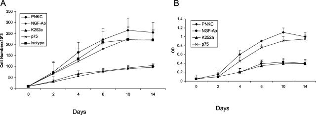
Nerve growth factor (NGF) influences the key pathological events of psoriasis: keratinocyte proliferation, angiogenesis, and T-cell activation. We have systematically examined the kinetics of NGF expression, keratinocyte proliferation, and migration of T lymphocytes in the epidermis in Koebner-induced developing psoriatic plaques. In skin traumatized by the tape-stripping method (n = 12), a marked up-regulation of NGF in Koebner-positive lesions (n = 7) was observed 24 hours after trauma. Synthesis of NGF reached its maximum level in the 2nd week. Furthermore, cultured keratinocytes from nonlesional skin of psoriasis patients produced 10 times higher levels of NGF compared with keratinocytes from healthy individuals. To substantiate the in vivo effect of NGF secreted by keratinocytes in psoriatic plaques, we studied psoriatic plaques and normal human skin in a SCID-human skin xenograft model. The transplanted psoriatic plaques demonstrated marked proliferation of NGF-R (p75)-positive nerve fibers compared with only a few nerves in the transplanted normal human skin. Our results demonstrate that 1) in a developing psoriatic lesion, up-regulation of NGF together with keratinocyte proliferation are early events and precede epidermotropism of T lymphocytes; 2) keratinocytes in patients with psoriasis are primed to produce elevated levels of NGF; and 3) NGF synthesized by these keratinocytes is functionally active.


Figures

Figure 1
Figure 1
A–G: NGF is increased in the keratinocytes of Koebner-positive lesions. Compared to nonlesional psoriasis skin (A), marked up-regulation of NGF is demonstrated in the keratinocytes within 24 hours of traumatization in the developing Koebner-positive lesion (B). C: Whereas after 24 hours of traumatization in a Koebner-negative lesion no significant up-regulation of NGF is noticed. D (2nd week), E (3rd week), and F (4th week) in a developing Koebner-positive psoriasis lesion demonstrate that intracellular expression of NGF remained high as the lesion continued to develop into a mature psoriasis plaque. Representative positively stained NGF cells are marked with the arrows. G: Immunocytochemical control for the primary antibody: section from the same tissue of F stained without primary anti-NGF antibody does not show any positively stained NGF cell.
Figure 2
Figure 2
A–C: Increased NGF-R-positive nerve fibers in Koebner-positive lesions. In parallel with the up-regulation of lesional NGF (Figure 1) marked proliferation of terminal cutaneous nerves with increased expression of NGF-R is noticed in the developing lesions of psoriasis. Figure 1B demonstrates sprouting of nerve fibers in the 2nd week whereas proliferation of these fibers along the side of the rete ridges a unique feature of psoriatic plaques is demonstrated in C.
Figure 3
Figure 3
Kinetics of CD3+ T-lymphocyte infiltrates in Koebner-positive psoriatic lesion. Immunoperoxidase staining for CD3 infiltrates in developing lesions of psoriasis. Developing psoriasis lesion in the 1st week demonstrates epidermal thickening, early rete peg formation whereas scattered trafficking of CD3-positive T lymphocytes are localized perivascularly in the dermis. In the 2nd week CD3 infiltrates migrate to dermo-epidermal junction, whereas significant epidermotropisim of CD3+ lymphocytes could be seen in the 3rd week.
Figure 4
Figure 4
Clinical features of transplanted psoriatic plaque in SCID mouse. Representative transplanted psoriatic plaque in SCID mouse, 4 weeks after transplantation. This plaque shows typical features of psoriasis-like scaling, thickening of the skin, and erythema.
Figure 5
Figure 5
Histological features of transplanted psoriatic plaque. A: H&E staining of a biopsy from a 4-week-old psoriatic plaque transplant. The figure shows typical histopathological features of psoriasis with marked acanthosis, infiltration of mononuclear cells in papillary dermis, and parakeratosis. B: A biopsy from transplanted plaque was stained for CD8+ T cells. White arrows indicate intraepidermal CD8+ T cells, an immunohistopathological hallmark of a psoriasis plaque.
Figure 6
Figure 6
Regeneration of nerve fiber in the transplanted normal human skin and psoriatic plaque. A: No significant regeneration of cutaneous nerves in the transplanted normal human skin. B: A biopsy from 4-day-old plaque transplant was stained for NGF-R with anti-NGF-R antibody. Note that there is no NGF-R-positive fiber in this section. C–E: Biopsies from psoriatic plaques after 4 weeks of transplantation show strongly positive staining for NGF-R-positive nerve fibers.
Figure 7
Figure 7
Psoriatic keratinocytes from nonlesional skin (PNKCs) produce more NGF than normal keratinocytes (NKCs). Ten thousand keratinocytes from psoriatic patients (n = 5) and normal controls (n = 5) were seeded into 35-mm-well tissue-culture plates. Two hundred μl of supernatant was collected every 24 hour from each culture. NGF level was detected by ELISA. Culture medium alone was taken as background control for ELISA. Each point represents the mean ± SD of five experiments and each experiment is done in triplicate.
Figure 8
Figure 8
A and B: Secreted NGF acting through its high-affinity receptor trkA induces an autocrine loop for keratinocyte proliferation in the cultured psoriatic keratinocytes from nonlesional skin (PNKCs). Ten thousand keratinocytes from psoriatic patients (n = 5) were seeded in 35-mm-well tissue-culture plates. Keratinocytes were cultured with medium only, K252a (100 nmol/L), NGF-neutralizing monoclonal antibody (100 ng/ml), p75 monoclonal antibody (100 ng/ml), isotype control (mouse IgG, 100 ng/ml) antibody. Keratinocyte proliferation at different time points was performed by the cell-counting method (A) and hexosaminidase assay (B). Keratinocyte cell numbers increased with duration of the culture period. Keratinocytes cultured in the presence of the NGF neutralizing antibody or K252a, the trkA inhibitor had significant growth inhibition; compared to no effect in presence of the anti-p75 monoclonal antibody that binds with the low-affinity NGF-R. Marked inhibition of keratinocyte proliferation by the anti-NGF neutralizing antibody and K252a suggests that secreted NGF acting through its high-affinity receptor trkA induces an autocrine loop for keratinocyte proliferation in the cultured PNKCs. Each test in each assay method was done in triplicate and repeated five times. Each point represents the mean ± SD.

Comment in

References

    1. Aloe L. Nerve growth factor and neuroimmune responses: basic and clinical observations. Arch Physiol Biochem. 2001;109:354–356. - PubMed
    1. Hattori A, Iwasaki S, Murase K, Tsujiimoto M, Sato M, Hyashi K, Kohno M. Tumor necrosis factor is markedly synergistic with interleukin1 and interferon gamma in stimulating the production of nerve growth factor in fibroblasts. FEBS Lett. 1994;340:177–180. - PubMed
    1. Heese K, Hock C, Otten U. Inflammatory signals induce neurotrophin expression in human microglial cells. J Neurochem. 1998;70:699–707. - PubMed
    1. Farber EM, Nickoloff BJ, Recht B, Fraki JE. Stress, symmetry, and psoriasis: possible role of neuropeptides. J Am Acad Dermatol. 1986;14:305–311. - PubMed
    1. Raychaudhuri SP, Farber EM. Are sensory nerves essential for the development of psoriasis lesions? J Am Acad Dermatol. 1993;28:488–489. - PubMed

MeSH terms