. 2008 Jun 1;111(11):5390-9.
doi: 10.1182/blood-2007-10-119743. Epub 2008 Mar 18.

EPO receptor circuits for primary erythroblast survival

Affiliations

EPO receptor circuits for primary erythroblast survival

Pradeep Sathyanarayana et al. Blood. .

Abstract

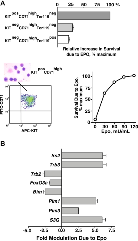
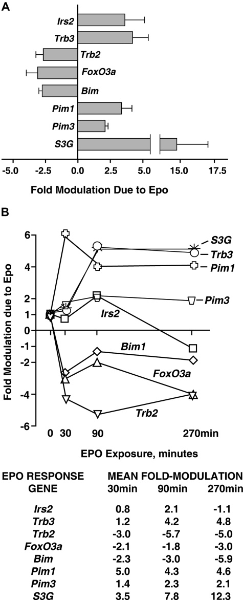
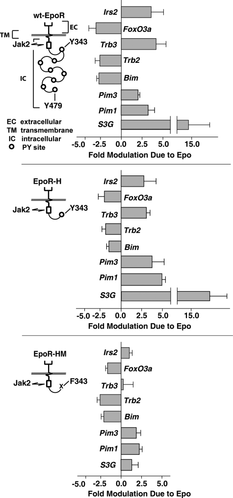
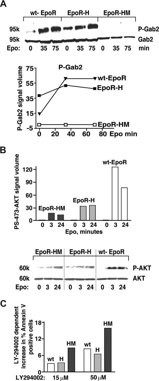
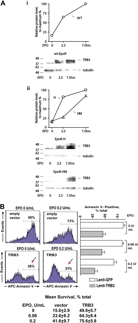
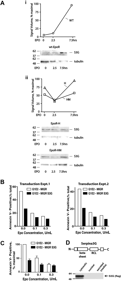
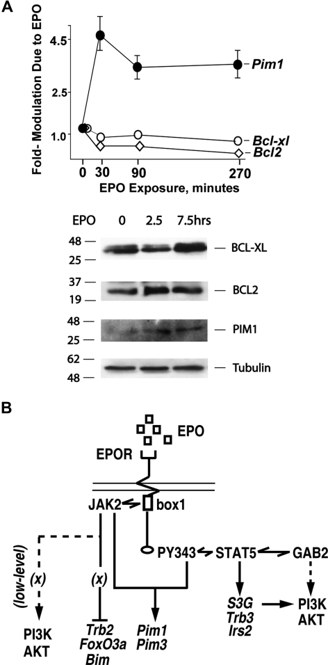
EPO functions primarily as an erythroblast survival factor, and its antiapoptotic actions have been proposed to involve predominantly PI3-kinase and BCL-X pathways. Presently, the nature of EPO-regulated survival genes has been investigated through transcriptome analyses of highly responsive, primary bone marrow erythroblasts. Two proapoptotic factors, Bim and FoxO3a, were rapidly repressed not only via the wild-type EPOR, but also by PY-deficient knocked-in EPOR alleles. In parallel, Pim1 and Pim3 kinases and Irs2 were induced. For this survival gene set, induction failed via a PY-null EPOR-HM allele, but was restored upon reconstitution of a PY343 STAT5-binding site within a related EPOR-H allele. Notably, EPOR-HM supports erythropoiesis at steady state but not during anemia, while EPOR-H exhibits near wild-type EPOR activities. EPOR-H and the wild-type EPOR (but not EPOR-HM) also markedly stimulated the expression of Trb3 pseudokinase, and intracellular serpin, Serpina-3G. For SERPINA-3G and TRB3, ectopic expression in EPO-dependent progenitors furthermore significantly inhibited apoptosis due to cytokine withdrawal. BCL-XL and BCL2 also were studied, but in highly responsive Kit(pos)CD71(high)Ter119(neg) erythroblasts, neither was EPO modulated. EPOR survival circuits therefore include the repression of Bim plus FoxO3a, and EPOR/PY343/STAT5-dependent stimulation of Pim1, Pim3, Irs2 plus Serpina-3G, and Trb3 as new antiapoptotic effectors.


Figures

Figure 1
Figure 1
Bone marrow erythroblast preparations, EPO-dependent survival responses, and transcriptome-based identification of EPO-regulated survival genes. (A) Bone marrow preparations were expanded for 3 days in SP34-EX medium to yield KitposCD71highTer119neg, KitnegCD71highTer119neg, and KitnegCD71highTer119pos erythroblast populations. Each was then purified by MACS and/or FACS, and increases in survival as afforded by EPO were determined. Specifically, cells were cultured in SP34-EX medium in the absence of SCF and presence of EPO at 0.1 U/mL. At 18 hours, frequencies of apoptotic cells were assayed using FITC–annexin-V and flow cytometry. Values are normalized means plus or minus SE. KitposCD71highTer119neg erythroblasts as purified by multiparameter MACS also were visualized in cytospin preparations and were assayed for protection against programmed cell death at a range of EPO doses. In the lower left panel, flow cytometry analyses also depict the homogeneity of isolated KitposCD71highTer119neg cells. (B) KitposCD71highTer119neg erythroblasts were cultured for 6 hours in the absence of hematopoietic cytokines, and were then exposed to EPO (± 5 U/mL) for 90 minutes. This included parallel processing of bone marrow–derived erythroblasts from n = 4 independent mice. After EPO exposure, RNA was isolated directly; 4 μg was used in biotin-cRNA syntheses and Affymetrix 430-2.0 array hybridizations. Among candidate (anti)apoptosis-related genes, 8 proved to be modulated by EPO at a confidence interval more than 99%. This included Irs2, Trb3, Trb2, Foxo3a, Bim, Pim1, Pim3, and Serpina3G (S3G). Values are mean-fold modulation plus or minus SD.
Figure 2
Figure 2
Quantitative RT-PCR analyses of EPO modulation of Irs2, Foxo3a, Trb2, Trb3, Bim, Pim1, Pim3, and S3G expression. (A) For each of these (anti)apoptotic factors, multifold modulation by EPO was confirmed by RT-QPCR within KitposCD71highTer119neg erythroblasts as exposed to EPO for 90 minutes. Values are means plus or minus SD for n = 3 independent samples. (B) For each factor, time courses of modulation by EPO also were analyzed. Here, KitposCD71highTer119neg erythroblasts were isolated, cultured for 6 hours in the absence of hematopoietic cytokines, and exposed to EPO (2.5 U/mL). At the indicated intervals, RNA was prepared, reverse-transcribed, and used in quantitative PCR analyses (vs beta-actin as a normalizing control). For an independent sample set, time course analyses were repeated. Results are illustrated for 2 such analyses as mean fold modulation due to EPO for each EPO-modulated candidate survival factor.
Figure 3
Figure 3
EPO induction of Irs2, Trb3, S3G, Pim1, and PIm3 depends upon EPOR-H PY343 signals, while EPO inhibition of Foxo3a, Trb2, and Bim is EPOR-PY independent. Erythroid progenitor cells were expanded (in SP34-EX medium) from bone marrow preparations for wt-EPOR, EPOR-H, and EPOR-HM mice (n = 3 independent mice per group). For wt-EPOR, EPOR-HM, and EPOR-H EPO receptor alleles, schematics are provided. KitposCD71highTer119neg erythroblasts then were isolated, cultured for 5.5 hours in the absence of hematopoietic cytokines, and exposed to EPO (± 2.5 U/mL). At 90 minutes, RNA was isolated. cDNA was then prepared, and levels of Irs2, Trb3 Trb2, Foxo3a, Bim, Pim1, Pim3, and S3G transcripts were determined by quantitative PCR. Values are mean-fold modulation plus or minus SD.
Figure 4
Figure 4
An EPOR-PY343 STAT5 axis mediates GAB2 but not AKT activation. (A) KitposCD71highTer119neg erythroblasts were prepared from wt-EPOR, EPOR-H, and EPOR-HM bone marrow cell expansion cultures. Cells were then cultured for 5.5 hours in IMDM containing transferrin (50 μg/mL), insulin (15 ng/mL), and BSA (0.5%). EPO (2 U/mL) was then added and at 0, 35, and 75 minutes lysates were prepared and levels of PY452 GAB2 and total GAB2 were determined by Western blotting. Outcomes were analyzed quantitatively by scanning densitometry (bottom panel). (B) Coupling to AKT is deficient for not only EPOR-HM but also EPOR-H alleles. Bone marrow–derived KitposCD71high erythroblasts were prepared and purified (as described in panel A) from EPOR-HM, EPOR-H, and wt-EPOR mice. Following withdrawal of hematopoietic cytokines (for 5.5 hours), cells were exposed to EPO (2.5 U/mL) and at the indicated intervals lysates were prepared. Levels of phospho-S473-AKT then were determined by Western blotting (and normalized for AKT levels/loading). Note the limited activation of AKT in not only EPOR-HM but also EPOR-H erythroblasts. Vertical lines indicate reassembled segments from a single, uniform en-hanced chemiluminescence (ECL) exposure. (C) Bone marrow–derived KitposCD71highTer119neg erythroblasts expressing an EPOR-H allele are efficiently protected by EPO against apoptosis, while EPOR-HM erythroblasts are not; KitposCD71high wt-EPOR, EPOR-H, and EPOR-HM erythroblasts were expanded and purified. Cells were then treated with LY274002 to inhibit PI3K,and were cultured in the presence of EPO at 0.2 U/mL. At 18 hours of culture, frequencies of apoptotic cells were determined by annexin-V staining and flow cytometry. Representative outcomes for LY274002 dosing at 15 and 50 μM are shown.
Figure 5
Figure 5
TRB3 induction via EPOR alleles, and TRB3-mediated survival effects in EPO-dependent erythroid progenitor cells. (A) KitposCD71highTer119neg erythroblasts were expanded, and purified from marrow progenitors as prepared from wt-EPOR, EPOR-H, and EPOR-HM mice. Cells then were cultured for 5 hours in 50 μg/mL transferrin, 0.1% BSA, 0.1 mM 2-MF, 15 ng/mL insulin, IMDM and subsequently exposed to EPO (2.5 U/mL). At the intervals indicated, cell lysates were prepared and analyzed for TRB3 expression via Western blotting. Outcomes for the wt-EPOR are illustrated in panel Ai, and for EPOR-HM and EPOR-H alleles in panel Aii. (B) TRB3 inhibits apoptosis due to cytokine withdrawal in EPO-dependent UT7epo cells. Lentiviruses encoding TRB3 or GFP only (empty control vector) were prepared and used to stably transduce UT7epo cells. GFPpos cells were then isolated by FACS, and possible effects of TRB3 on survival were assessed in the presence of EPO at limiting doses. As assayed via staining with APC–annexin-V, significant protection against apoptosis was afforded by TRB3. Representative flow cytometry data are shown (left panels) together with mean TRB3 survival effects (± SE, n = 3). Mean outcomes for 2 independent sets of experiments also are shown (botom panel).
Figure 6
Figure 6
EPO modulation of endogenous S3G expression in wt-EPOR, EPOR-H, and EPOR-HM erythroblasts, and S3G effects on erythroid progenitor cell survival. (A) KitposCD71highTer119neg erythroblasts were isolated from wt-EPOR, EPOR-H, and EPOR-HM bone marrow expansion cultures. Cells then were cultured for 5 hours in 50 μg/mL transferrin, 0.1% BSA, 0.1 mM 2-ME, 15 ng/mL insulin, IMDM. At the time intervals indicated, cell lysates were prepared and analyzed for S3G expression via Western blotting. Outcomes for the wt-EPOR are illustrated in panel Ai, and for EPOR-HM and EPO-H alleles in panel Aii. (B) Epo-dependent G1E/JC4 cells were transduced with a MIGR-S3G retroviral construct, or with an empty MIGR vector as a negative control. For G1E/JC4-S3G and G1E/JC4-MIGR cells in exponential growth phase, EPO was withdrawn for 6 hours and subsequently was provided at the doses indicated. At 24 hours, frequencies of apoptotic cells were assayed by staining with annexin-V and flow cytometry. Transduction sets nos. 1 and 2 represent independently transduced cell populations. (C) Average effects of S3G on G1E/JC4 cell survival for 4 independent analyses also are illustrated. Values are normalized means plus or minus SE. (D) Via Western blotting of cellular fractions and concentrated media, ectopically expressed S3G was observed to localize to a cytosolic fraction. S3G's basic structure, including its serpin domain and reactive center loop (RCL), also is diagrammed.
Figure 7
Figure 7
EPO-independent activation of BCL-XL and BCL2 expression at discrete stages of (pro)erythroblast development. (A) In primary bone marrow erythroblasts, neither BCL-XL nor BCL2 expression is EPO-modulated. KitposCD71highTer119neg erythroblasts were expanded from wt-EPOR bone marrow preparations. Cells then were cultured for 5 hours in 50 μg/mL transferrin, 0.1% BSA, 15 ng/mL insulin, 0.1 mM 2-ME, IMDM. EPO was then added (2.5 U/mL), and at 0, 2.5, and 7.5 hours cell lysates were prepared. Lysates then were analyzed by Western blotting for levels of BCL-XL, BCL2, PIM1, and beta-tubulin. In parallel, possible EPO modulation of Bcl-x or Bcl2 transcripts also was analyzed, here at 0, 30, 90, and 270 minutes of EPO exposure. Values are mean-fold modulation plus or minus SE. (B) Mapping of EPOR subdomains to EPO-regulated survival factor circuits. Presently defined EPO-modulated transcriptional response circuits are outlined. An EPOR JAK2-only circuit mediates Foxo3a, Bim, and Trb2 repression. In parallel, an EPOR/PY343/STAT5 axis enhances Pim1 and Pim3 expression, and affords EPO induction of Irs2, S3G, and Trb3.

Similar articles

Cited by

References

    1. Fisher JW, Koury S, Ducey T, Mendel S. Erythropoietin production by interstitial cells of hypoxic monkey kidneys. Br J Haematol. 1996;95:27–32. - PubMed
    1. Rankin EB, Biju MP, Liu Q, et al. Hypoxia-inducible factor-2 (HIF-2) regulates hepatic erythropoietin in vivo. J Clin Invest. 2007;117:1068–1077. - PMC - PubMed
    1. Wu H, Liu X, Jaenisch R, Lodish HF. Generation of committed erythroid BFU-E and CFU-E progenitors does not require erythropoietin or the erythropoietin receptor. Cell. 1995;83:59–67. - PubMed
    1. Koury MJ, Bondurant MC. Erythropoietin retards DNA breakdown and prevents programmed death in erythroid progenitor cells. Science. 1990;248:378–381. - PubMed
    1. Jelkmann W. Erythropoietin after a century of research: younger than ever. Eur J Haematol. 2007;78:183–205. - PubMed

Publication types

Substances