. 2008 Apr 8;98(7):1274-84.
doi: 10.1038/sj.bjc.6604286. Epub 2008 Mar 18.

Involvement of focal adhesion kinase in cellular invasion of head and neck squamous cell carcinomas via regulation of MMP-2 expression

Affiliations

Involvement of focal adhesion kinase in cellular invasion of head and neck squamous cell carcinomas via regulation of MMP-2 expression

M Canel et al. Br J Cancer. .

Abstract

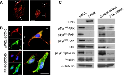
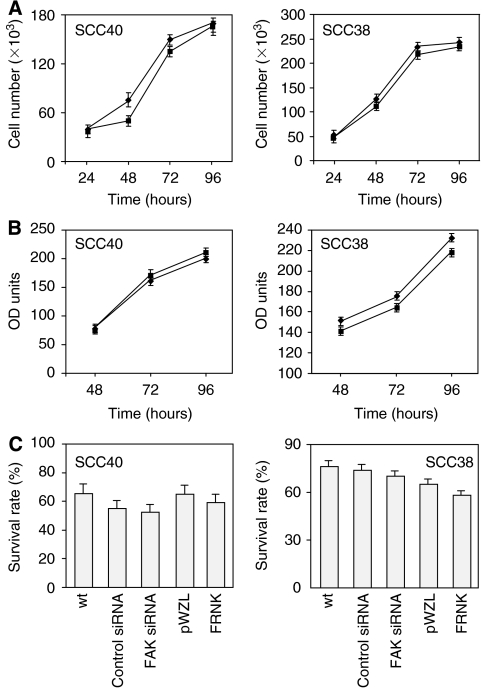
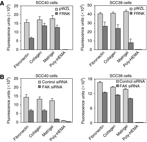
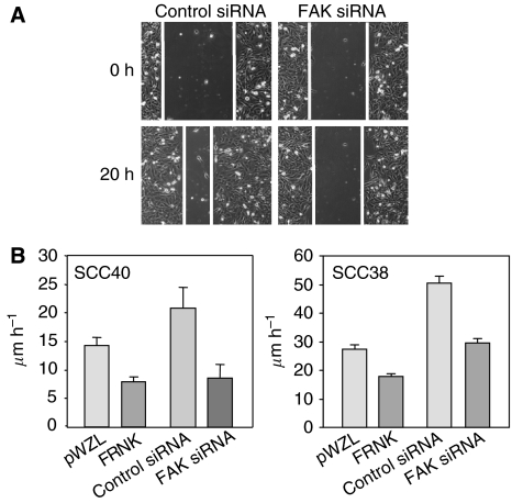
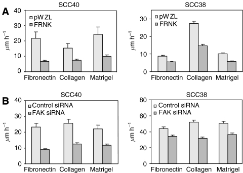
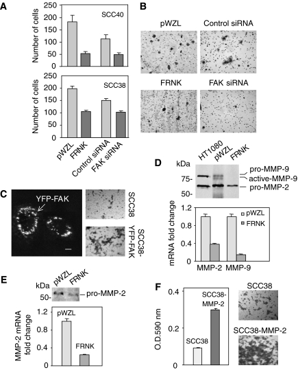
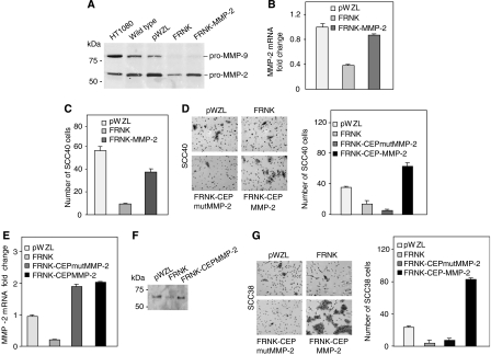
Focal adhesion kinase (FAK) is considered intimately involved in cancer progression. Our previous research has demonstrated that overexpression of FAK is an early and frequent event in squamous cell carcinomas of the supraglottic larynx, and it is associated with the presence of metastases in cervical lymph nodes. The purpose of this study was to examine the functional role of FAK in the progression of head and neck squamous cell carcinomas (HNSCC). To this end, expression of FAK-related nonkinase (FRNK) or small interfering RNA (siRNA) against FAK was used to disrupt the FAK-induced signal transduction pathways in the HNSCC-derived SCC40 and SCC38 cell lines. Similar phenotypic effects were observed with the two methodological approaches in both cell lines. Decreased cell attachment, motility and invasion were induced by FRNK and FAK siRNA, whereas cell proliferation was not impaired. In addition, increased cell invasion was observed upon FAK overexpression in SCC cells. FRNK expression resulted in a downregulation of MMP-2 and MMP-9 expression. Interestingly, MMP-2 overexpression in FRNK-expressing cells rescued FRNK inhibition of cell invasion. This is the first demonstration of a direct rescue of impaired cell invasion by the re-expression of MMP-2 in a tumour cell type with decreased expression of functional FAK. Collectively, these data reported here support the conclusion that FAK enhances invasion of HNSCC by promoting both increased cell motility and MMP-2 production, thus providing new insights into possible therapeutic intervention strategies.


Figures

Figure 1
Figure 1
Inhibition of FAK-mediated signalling by expression of FRNK or siRNA against FAK in HNSCC-derived SCC40 cells. (A) Confocal microscopy images of SCC40 cells transiently transfected with pEGFP-FRNK and stained with phalloidin-TRITC to visualise actin organisation (GFP-FRNK, green fluorescence; phalloidin-TRITC, red fluorescence). Images show transfected cells as well as nontransfected cells (marked with arrows), which served as internal controls. As shown, FRNK is localised to focal contacts, docking sites of actin cytoskeleton to the extracellular matrix. Scale bars, 10 μm. (B) Confocal microscopy images of SCC40 cells stably transfected with pWZL vector alone or pWZL-FRNK (FAK, green fluorescence; vinculin, red fluorescence). Images show that FAK colocalise with vinculin at focal contact sites (marked with arrows) in pWZL-SCC40 cells whereas is mainly detected in the cytoplasm in FRNK-SCC40 cells. Scale bars, 20 μm (pWZL-SCC40) and 15 μM (FRNK-SCC40). (C) Western blot analysis showing expression of FRNK, pTyr397-FAK, pTyr861-FAK, pTyr925-FAK, total FAK, pTyr118-paxillin and total paxillin in SCC40 cells stably transfected with pWZL-FRNK (FRNK) or treated with FAK-siRNA. Cells infected with empty pWZL vector (pWZL) or transfected with control siRNA served as the control. The membranes were stripped and reprobed with anti-α-tubulin antibody to assure even loading of proteins in each lane.
Figure 2
Figure 2
FAK inhibition does not affect cell proliferation or survival in SCC cells. (A) Cell proliferation of pWZL-(♦) and FRNK-SCC cells (▪). Cells (50 × 103) were plated and 24, 48, 72, or 96 h later the number of cells was measured by direct counting of trypan blue-excluding cells using a hemacytometer. Values are mean±s.d. from a representative experiment performed in triplicate in a series of three. (B) MTS assays were performed with pWZL-(♦) and FRNK-SCC cells (▪) growth for 48, 72 and 96 h. Values are mean±s.d. from a representative experiment performed in triplicate in a series of three. (C) Survival rate of FRNK- or pWZL-SCC cells and SCC cells treated with FAK- or control-siRNA. The fraction of viable cells was measured by using the FITC-Annexin V apoptosis detection kit. Values are mean of average±s.d. from three independent experiments done in triplicate.
Figure 3
Figure 3
Inhibition of FAK expression or activity impaired cell attachment in SCC40 and SCC38 cells. Cell adhesion on different substrates was determined as described in Material and Methods in SCC40 and SCC38 cells stably transfected with pWZL-FRNK (A) and cells treated with FAK-siRNA (B) and compared with that measured in their corresponding control cells (pWZL and control siRNA). Values are mean of average±s.d. from four independent experiments done in triplicate.
Figure 4
Figure 4
Inhibition of FAK-mediated signalling pathway attenuates cell migration in SCC40 and SCC38 cells. Wound healing assays were performed in FRNK- and pWZL-expressing SCC40 and SCC38 cells, and cells treated with FAK- or control-siRNA. (A) Representative images captured with a × 10 objective at the time of wounding (0 h) or 20 h after wounding (20 h) in SCC40 cells transfected with control- or FAK-siRNA. (B) Rate of front migration of cell monolayers analysed by time-lapse video microscopy. At least 15 different fields were randomly chosen across the wound length. Values are mean of average±s.d. from three independent experiments.
Figure 5
Figure 5
Inhibition of FAK-mediated signalling inhibits cell motility in SCC40 and SCC38 cells. FRNK- or pWZL-SCC40 and SCC38 cells (A) and cells treated with FAK- or control-siRNA (B) were plated and images taken every 15 min over a 12-h period. Quantification of linear cell motility was determined by tracing the single cells using the cell-tracking software (Kenetic Imaging Ltd). About 100 individual cells per cell line were analysed. Values are mean of average±s.d. from three independent experiments done in triplicate.
Figure 6
Figure 6
Inhibition of FAK-mediated signalling decreases cellular invasion through Matrigel and MMP-2 expression in SCC cells. (A and B) FRNK- or pWZL-SCC cells and SCC cells treated with FAK- or control-siRNA were seeded in serum-free media in the upper chamber of Matrigel transwells. The lower chamber was loaded with regular media supplemented with 10% fetal bovine serum and 5% BSA. After 24 h at 37°C in 5% CO2, the top filter was scraped, and invading cells were fixed and stained. (A) All invading cells were counted under × 10 magnification. Values are mean of average±s.d. from a representative experiment in a series of three done in triplicate. (B) Representative images from three separate experiments performed in SCC40 cells. (C) SCC38 cells were transiently transfected with either pYFP-FAK or vector alone. Picture on the left is a representative confocal microscope image showing that YFP-FAK fusion protein localises at focal sites. Scale bars, 10 μm. Matrigel invasion assays were performed 24 h after transfection. Representative images from three separate matrigel invasion experiments performed in SCC38 cells transfected with either vector alone or pYFP-FAK are shown on the right. (D and E), FRNK- or pWZL-SCC40 (D) and FRNK- or pWZL-SCC38 (E) cells were cultured in serum-free media. After 24 h, conditioned media and cells were harvested for gelatinase zymography and MMPs mRNA quantification, respectively. HT-1080 fibrosarcoma cells conditioned medium was used as positive control for zymography and migration standards. MMP-2 and MMP-9 transcripts were quantified using quantitative RT–PCR. The mean of relative expression to cyclophilin A housekeeping gene of at least three independent experiments is shown. (F) Matrigel invasion assay was performed in SCC38 cells 24 h after transfection with either pcDNA3-MMP-2 or vector alone. Cell staining with crystal violet and subsequent extraction with dimethylsulphoxide followed by spectrophotometry at 590 nm was performed for quantification of invading cells. All analyses were performed in triplicate. Representative images from three separate experiments are shown.
Figure 7
Figure 7
MMP-2 overexpression rescues FRNK inhibition of cell invasion in SCC40 (AD) and SCC38 (EG) cells. Gelatinase zymography (A), MMP-2 mRNA quantification (B) and in vitro invasive assays (C) were performed in FRNK-, pWZL-, and FRNK-MMP-2-SCC40 cells as indicated in Figure 6. (D) In vitro invasive assays performed in pWZL-, FRNK-, FRNK-CEP-MMP-2- and FRNK-CEPmutMMP-2-SCC40 cells. All invading cells were counted under × 10 magnification. Representative images from three separate experiments are shown. MMP-2 mRNA quantification (E), gelatinase zymography (F) and matrigel invasion assays (G) were performed in pWZL-, FRNK-, FRNK-CEP-MMP-2- and FRNK-CEPmutMMP-2-SCC38 cells as indicated above. Representative images of matrigel invasion assays in SCC38 cells are shown in (G). Experiments were repeated at least three times.

References

    1. Aguirre Ghiso JA (2002) Inhibition of FAK signaling activated by urokinase receptor induces dormancy in human carcinoma cells in vivo. Oncogene 21: 2513–2524 - PubMed
    1. Bellis SL, Miller JT, Turner CE (1995) Characterization of tyrosine phosphorylation of paxillin in vitro by focal adhesion kinase. J Biol Chem 270: 17437–17441 - PubMed
    1. Brunton VG, Avizienyte E, Fincham VJ, Serrels B, Metcalf III CA, Sawyer TK, Frame MC (2005) Identification of Src-specific phosphorylation site on focal adhesion kinase: dissection of the role of Src SH2 and catalytic functions and their consequences for tumor cell behavior. Cancer Res 65: 1335–1342 - PubMed
    1. Cance WG, Harris JE, Iacocca MV, Roche E, Yang X, Chang J, Simkins S, Xu L (2000) Immunohistochemical analyses of focal adhesion kinase expression in benign and malignant human breast and colon tissues: correlation with preinvasive and invasive phenotypes. Clin Cancer Res 6: 2417–2423 - PubMed
    1. Canel M, Secades P, Rodrigo JP, Cabanillas R, Herrero A, Suarez C, Chiara MD (2006) Overexpression of focal adhesion kinase in head and neck squamous cell carcinoma is independent of fak gene copy number. Clin Cancer Res 12: 3272–3279 - PubMed

Publication types

MeSH terms

Substances