. 2008 May;5(5):280-90.
doi: 10.1038/ncponc1089. Epub 2008 Mar 18.

Mechanisms of disease: epithelial-mesenchymal transition--does cellular plasticity fuel neoplastic progression?

Affiliations

Mechanisms of disease: epithelial-mesenchymal transition--does cellular plasticity fuel neoplastic progression?

Eva A Turley et al. Nat Clin Pract Oncol. 2008 May.

Abstract

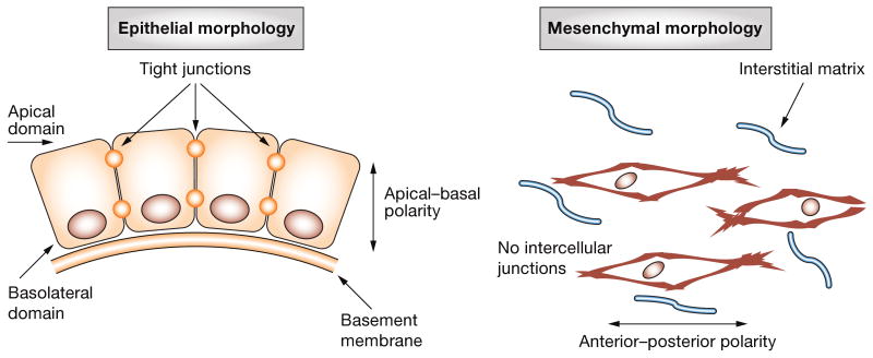
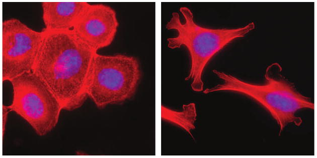
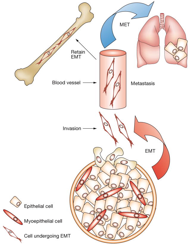
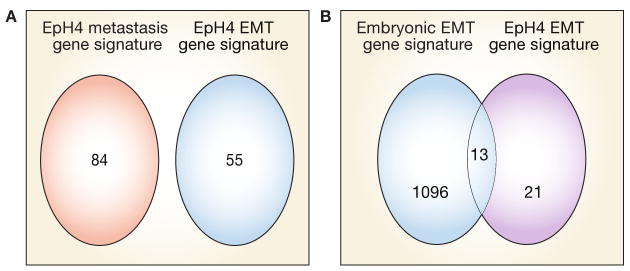
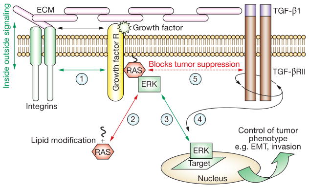
Epithelial-mesenchymal transition (EMT) is a phenotypic conversion that facilitates organ morphogenesis and tissue remodeling in physiological processes, such as embryonic development and wound healing. A similar phenotypic conversion is also detected in fibrotic diseases and neoplasia, and is associated with disease progression. EMT in cancer epithelial cells often seems to be an incomplete and bidirectional process. In this Review, we discuss the phenomenon of EMT as it pertains to tumor development, focusing on exceptions to the commonly held rule that EMT promotes invasion and metastasis. We also highlight the role of RAS-controlled signaling mediators, ERK1, ERK2 and phosphatidylinositol 3-kinase, as microenvironmental responsive regulators of EMT.


Conflict of interest statement

Competing interests: The authors declared no competing interests.

Figures

Figure 1
Figure 1
Common morphologic characteristics of epithelial and mesenchymal cells. Epithelial morphology is characterized by an apical–basal polarity, contact with a basal basement membrane and formation of extensive cell–cell contacts, including tight junctions. An anterior–posterior polarity is lost if any cell–cell junctions and residency within a more unstructured interstitial matrix characterize mesenchymal morphology.
Figure 2
Figure 2
EMT of mammary epithelial cells. Treatment of mouse mammary epithelial cells with MMP3 stimulates breakdown of the epithelial structure and acquisition of a mesenchymal morphology. Red staining, f-actin; blue staining, DAPI. Abbreviations: DAPI, 4′-6-diamidino-2-phenylindole; EMT, epithelial–mesenchymal transition; MMP, matrix metalloproteinase.
Figure 3
Figure 3
The dynamic role of EMT in mammary gland neoplastic processes. EMT during mammary tumor progression is postulated to facilitate invasion and colonization of distant tissues by tumor cells. EMT enables efficient penetration of vessels and escape into distant tissues, such as the lung or bone. A mesenchymal phenotype might be retained or revert to an epithelial phenotype (MET), depending on the tissue microenvironment. For example, some microenvironments, such as those provided by bones, can offer selective growth for a mesenchymal phenotype, whereas others (e.g. lung) might favor growth of an epithelial phenotype. Abbreviations: EMT, epithelial–mesenchymal transition; MET, mesenchymal–epithelial transition.
Figure 4
Figure 4
Overlap between the EMT gene signature of EpH4 mammary cells and that of the embryonic palate. (A) A Venn diagram illustrating the overlap between the gene signature of EpH4 metastasis and the gene signature of EMT in EpH4. Both upregulated and downregulated genes are represented. These results show that EMT can be distinguished from metastasis as a molecular process. (B) A Venn diagram showing the number of upregulated EMT-specific genes in EpH4 cells that are also upregulated or downregulated by at least twofold in the embryonic palate undergoing EMT. Both EMT processes are in response to TGF-β1. Approximately 50% of EMT-specific genes upregulated in EpH4 cells are also upregulated during EMT associated with embryonic palate morphogenesis. Based on data from Jechlinger M et al. (2003) Oncogene 22: 7155–7169, and LaGamba D et al. (2005) Dev Dyn 234: 132–142. Abbreviation: EMT, epithelial–mesenchymal transition.
Figure 5
Figure 5
Microenvironmental and spatial regulation of signaling pathways controlling EMT. The RAS–ERK1/ERK2 pathway is an example of a signaling module that is responsive to microenvironmental cues and requires specific subcellular localization to determine the consequences of gene expression and tumor phenotype. A simplified version of this complex process is illustrated in the diagram. ECM components interact with integrin receptors at the cell surface (step 1) and the affinity of this interaction is modified by growth-factor-regulated signaling, in a process known as ‘inside-out signaling’ (steps 1–3). Conversely, the affinity of the integrin–ECM interaction affects growth-factor-regulated signaling, in a process known as ‘outside-in signaling’ (steps 2–3). The collective interactions between integrins and growth factor receptors promote the localization and activation of lipid-modified RAS (step 2) at the inner cell membrane leaflet (step 4). Activated RAS then selectively activates kinases, such as ERK1/ERK2 and other pathways, such as the PI3K/AKT pathway. RAS also blocks the tumor-suppressing activity of TGF-β1 by linking this pathway to ERK1/ERK2 signaling pathways (step 5). This linkage promotes the proinvasion properties of TGF-β1. Activated ERK1/ERK2 must translocate to the nucleus or cell-adhesion sites (known as ‘focal contacts’) to have access to target proteins that regulate EMT, motility and/or invasion. ERK1-regulated and ERK2-regulated gene expression (e.g. MMP9) further modifies the microenvironment of the tumor cell, thereby affecting the activation status of the integrin–growth factor receptor signaling pathway. This downstream consequence of RAS–ERK1–ERK2 activation and the reversal of steps 1–5 have profound effects on the phenotype even if other pathways are mutated. Abbreviations: AKT, protein kinase B; ECM, extracellular matrix; EMT, epithelial–mesenchymal transition; ERK1, extracellular signal-regulated kinase 1; ERK2, extracellular signal-regulated kinase 2; MMP, matrix metalloproteinase; PI3K, phosphatidylinoinositol 3-kinase; TGF, transforming growth factor.

References

    1. Lee JM, et al. The epithelial-mesenchymal transition: new insights in signaling, development, and disease. J Cell Biol. 2006;172:973–981. - PMC - PubMed
    1. Valles AM, et al. Acidic fibroblast growth factor is a modulator of epithelial plasticity in a rat bladder carcinoma cell line. Proc Natl Acad Sci USA. 1990;87:1124–1128. - PMC - PubMed
    1. Kalluri R, Neilson EG. Epithelial-mesenchymal transition and its implications for fibrosis. J Clin Invest. 2003;112:1776–1784. - PMC - PubMed
    1. Desmouliere A, et al. The stroma reaction myofibroblast: a key player in the control of tumor cell behavior. Int J Dev Biol. 2004;48:509–517. - PubMed
    1. Nuyten DS, et al. Predicting a local recurrence after breast-conserving therapy by gene expression profiling. Breast Cancer Res. 2006;8:R62. - PMC - PubMed