. 2008 Mar 19;3(3):e1808.
doi: 10.1371/journal.pone.0001808.

A gamma-herpesvirus glycoprotein complex manipulates actin to promote viral spread

Affiliations

A gamma-herpesvirus glycoprotein complex manipulates actin to promote viral spread

Michael B Gill et al. PLoS One. .

Abstract

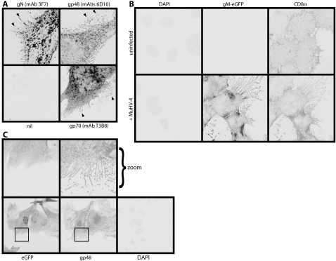
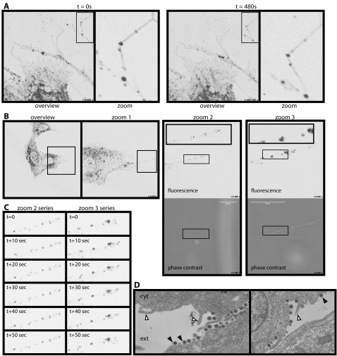
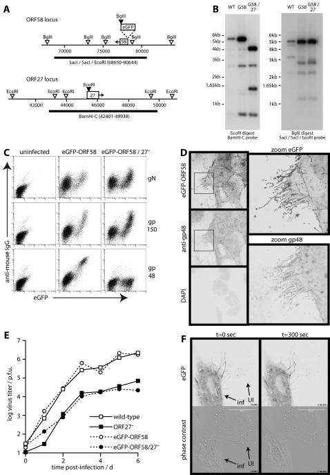
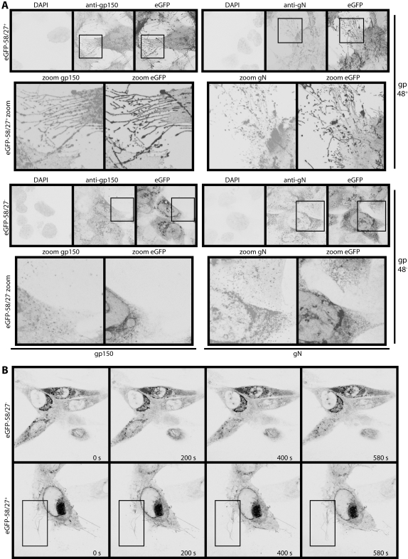
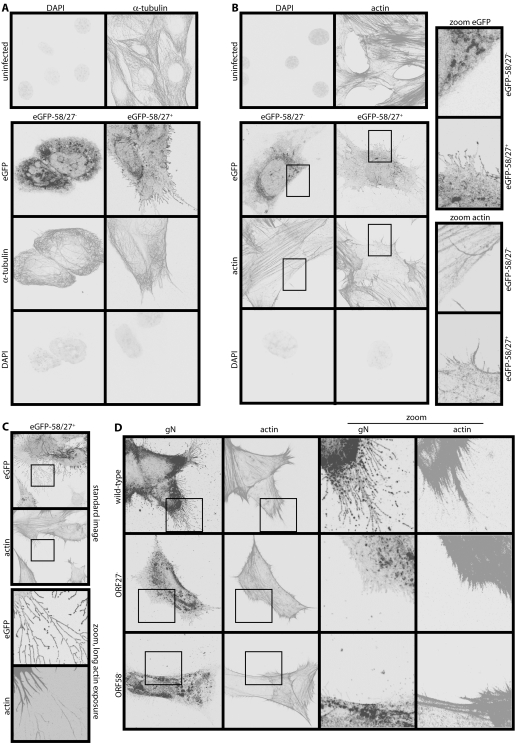
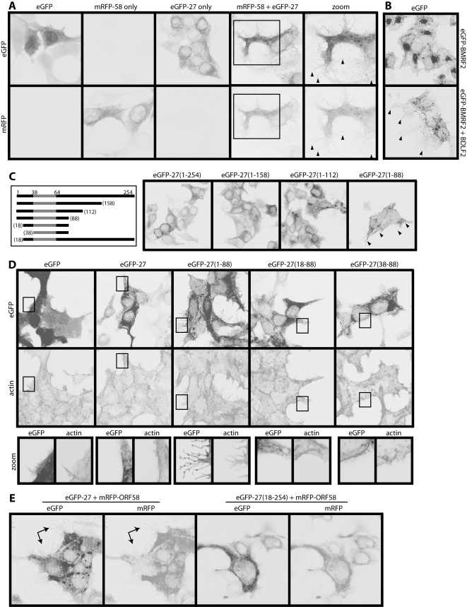
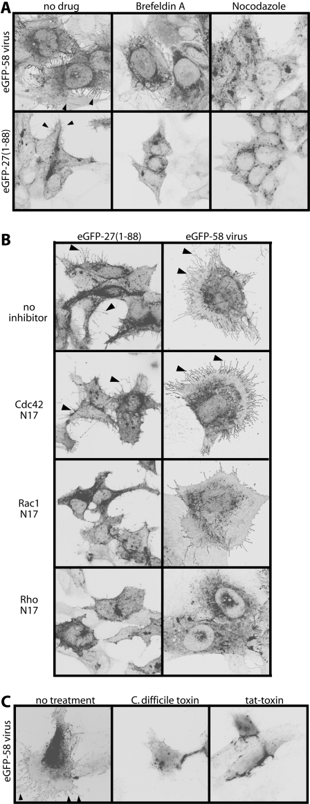
Viruses lack self-propulsion. To move in multi-cellular hosts they must therefore manipulate infected cells. Herpesviruses provide an archetype for many aspects of host manipulation, but only for alpha-herpesviruses in is there much information about they move. Other herpesviruses are not necessarily the same. Here we show that Murine gamma-herpesvirus-68 (MHV-68) induces the outgrowth of long, branched plasma membrane fronds to create an intercellular network for virion traffic. The fronds were actin-based and RhoA-dependent. Time-lapse imaging showed that the infected cell surface became highly motile and that virions moved on the fronds. This plasma membrane remodelling was driven by the cytoplasmic tail of gp48, a MHV-68 glycoprotein previously implicated in intercellular viral spread. The MHV-68 ORF58 was also required, but its role was simply transporting gp48 to the plasma membrane, since a gp48 mutant exported without ORF58 did not require ORF58 to form membrane fronds either. Together, gp48/ORF58 were sufficient to induce fronds in transfected cells, as were the homologous BDLF2/BMRF2 of Epstein-Barr virus. Gp48/ORF58 therefore represents a conserved module by which gamma-herpesviruses rearrange cellular actin to increase intercellular contacts and thereby promote their spread.


Conflict of interest statement

Competing Interests: The authors have declared that no competing interests exist.

Figures

Figure 1
Figure 1. Plasma membrane remodelling by MHV-68.
A. NIH-3T3 cells were infected (1 p.f.u./cell, 16 h) with wild-type MHV-68, then fixed, permeabilized and stained for viral glycoproteins. Positive staining appears black or grey. gN, gp48 and gp70 could each be seen on frond-like extensions of the infected cell plasma membrane (arrows). nil = secondary antibody only. B. NIH-3T3 cells stably expressing human CD8α were infected (1 p.f.u./cell, 16 h) or not with MHV-68 expressing eGFP-tagged gM. The cells were then fixed, permeabilized and stained for CD8α. Nuclei were counter-stained with DAPI. eGFP fluorescence was visualized directly. A similar MHV-68 induction of membrane fronds was seen using a lipophilic dye to stain the plasma membrane (data not shown). C. NIH-3T3 cells were infected (1 p.f.u./cell, 16 h) with MHV-68 expressing free eGFP, then fixed and stained for gp48 with mAb T8H3. eGFP fluorescence was visualized directly. Nuclei were counter-stained with DAPI. The zoomed images correspond to the boxed regions in the upper panels.
Figure 2
Figure 2. Relationship between membrane fronds and virions.
A. BHK-21 cells infected with MHV-68 expressing eGFP-tagged gM (1 p.f.u./cell, 16 h) were examined by time-lapse confocal microscopy. The eGFP signal appears as black/gray. Each zoomed image corresponds to the boxed region of the corresponding overview. The punctate fluorescence of distal membrane fronds is seen to change with time. See also Movies S1 and S2. B. BHK-21 cells were infected with gM-eGFP-tagged MHV-68 as in A. Time-lapse imaging then focussed on a single membrane process. Zoom 1 is the boxed region in the overview; zoom 2 is the boxed region of zoom 1, with its boxed region shown as a zoomed inset. Zoom 3 shows a region equivalent to the central part of zoom 2 at ×5 greater magnification. Again, its boxed region is shown as a further zoomed inset. The complete sets of zoom 2 and zoom 3 pictures make up Movies S3 and S4. C. Stills from Movies S3 and S4, which correspond to the zoom 2 and zoom 3 images in B, show the variation in eGFP+ dot distribution at 10sec intervals. D. BHK-21 cells were infected with MHV-68 (1 p.f.u./cell, 16h) then fixed and processed for transmission electron microscopy. cyt = cytoplasm; ext = extracellular. Closed arrows show virions. Open arrows show the bases of membrane fronds.
Figure 3
Figure 3. eGFP-tagged ORF58 decorates the membrane fronds.
A. Schematic diagram of the MHV-68 ORF58 and ORF27 loci. Filled arrowheads show restriction sites created by eGFP tagging ORF58 or disrupting ORF27. Open arrowheads show genomic restriction sites. The thick lines show probe locations. B. Southern blots of wild-type (WT), eGFP-tagged ORF58 (G58) and ORF27-deficient eGFP-tagged ORF28 (G58/27) viruses. The EcoRI digest plus BamHI-C probe gave predicted wild-type and G58 virus fragments of 5611bp, 2703bp, 744bp and 1342bp (difficult to see because of limited overlap with the probe). The EcoRI-restricted oligonucleotide introduced into ORF27 at genomic co-ordinate 45480 converted the 5611bp band to 4075bp plus 1536bp. The BglII digest plus SacI/SacI/EcoRI probe gave predicted WT fragments of 5300bp, 5165bp, 2294bp and 3379bp. The eGFP insertion just upstream of ORF58 converted the 3379 band to 1193bp+2916bp. C. BHK-21 cells were left uninfected or infected (0.5 p.f.u./cell, 16 h) with ORF27+ or ORF27 versions of the eGFP-ORF58 virus. The cells were then stained for gN (mAb 3F7), gp150 (mAb T1A1) or gp48 (mAb T8H3), each with phycoerythrin-conjugated goat anti-mouse IgG pAb. The data are from 1 of 3 equivalent experiments. D. NIH-3T3 cells were infected with eGFP-ORF58 MHV-68 (1 p.f.u./cell, 16 h), then fixed, permeabilized and stained for gp48 with mAb 6D10 plus Alexa568-conjugated goat anti-mouse IgG pAb. EGFP fluorescence was visualized directly. Nuclei were counterstained with DAPI. The zoomed images correspond to the boxed regions in the left-hand panels. The data are from 1 of 5 equivalent experiments. E. BHK-21 cells were infected (0.01 p.f.u./cell) with ORF27+ or ORF27 versions of either untagged (wild-type) or eGFP-ORF58-tagged MHV-68. Virus titers were determined by plaque assay at the times indicated. F. NIH-3T3 cells were infected with eGFP-ORF58 MHV-68 (1 p.f.u./cell, 16h) then examined by time-lapse confocal microscopy. An infected (inf) cell is shown next to an uninfected (UI) or only recently infected neighbour to demonstrate eGFP+ fronds reaching from one to the other. Movie S5 shows the difference between these cells in shape and motility. The findings were typical of >100 cells examined.
Figure 4
Figure 4. MHV-68-induced membrane changes are gp48-dependent.
A. NIH-3T3 cells were infected (1 p.f.u./cell, 16 h) with gp48+ (upper set of images) or gp48 (lower set of images) versions of eGFP-ORF58 MHV-68, then fixed, permeabilized and stained for gp150 (mAb T1A1, left set of images) or gN (mAb 3F7, right set of images). Each mAb was visualized with Alexa568-conjugated goat anti-mouse IgG pAb, eGFP was visualized directly, and nuclei were counterstained with DAPI. The zoomed images correspond to each boxed region above. B. NIH-3T3 cells were infected with ORF27 or ORF27+ eGFP-ORF58 viruses as in A, then visualized by time-lapse confocal microscopy. The boxed region highlights eGFP+ membrane fronds growing away from the ORF27+ infected cell surface. Times are indicated in each panel. The complete image sets make up Movies S6 and S7.
Figure 5
Figure 5. MHV-68-induced membrane fronds contain actin but not tubulin.
A. NIH-3T3 cells were left uninfected or infected (1 p.f.u./cell, 16 h) with either ORF27 or ORF27+ eGFP-ORF58 MHV-68. They were then fixed, permeabilized and stained for α-tubulin. EGFP fluorescence was visualized directly. Nuclei were counterstained with DAPI. B. NIH-3T3 cells were infected or not as in A, then fixed, permeabilized and stained for actin with Alexa568-conjugated phalloidin. EGFP fluorescence was visualized directly and nuclei were counterstained with DAPI. The zoomed images correspond to the boxed regions. They show coincident membrane fronds and actin spikes with ORF27+ infection and neither with ORF27 infection. C. NIH-3T3 cells were infected (1 p.f.u./cell, 16 h) with ORF27+ eGFP-ORF58-tagged MHV-68 and stained for actin as in B, but with longer exposure times for the zoomed images (which correspond to the boxed regions above) to show the very fine actin cores of the distal membrane fronds. D. NIH-3T3 cells were infected (1 p.f.u./cell, 16 h) with untagged wild-type, ORF27 or ORF58 MHV-68. The cells were then fixed, permeabilized and stained for gN with mAb 3F7 plus Alexa488-conjugated goat anti-mouse IgG pAb, and for actin with Alexa568-conjugated phalloidin.
Figure 6
Figure 6. ORF27 and ORF58 are sufficient to induce membrane fronds.
A. 293T cells were transfected with expression plasmids for eGFP, mRFP-tagged ORF58 (mRFP-58), eGFP-tagged ORF27 (eGFP-27), or mRFP-58 plus eGFP-27. Red and green fluorescence signals were examined 48 h later. The zoomed images match the boxed region in the mRFP-58+eGFP-27 transfection. Arrowheads show membrane fronds. B. 293T cells were transfected with an expression plasmid for eGFP-tagged BMRF2 (EBV ORF58 homolog), with or without an expression plasmid for BDLF2 (EBV ORF27 homolog). 48 h later, the eGFP signal was visualized. Arrowheads show membrane fronds, which were not seen with either plasmid alone. Note also eGFP-BMRF2 redistribution following BDLF2 transfection, much like that seen with ORFs 27 and 58. C. ORF27 truncations, each tagged with eGFP (our mAbs only recognize full-length ORF27) were transfected into 293T cells. 48 h later, the cells were fixed and eGFP fluorescence visualized. The numbers correspond to amino acid residues of the full-length protein (gp48). The gp48 C-terminal domain is extracellular. The shaded region corresponds to its transmembrane domain. The arrows show membrane fronds, which were prominent only with the eGFP-27(1-88) mutant. Note also the redistribution of eGFP fluorescence. D. 293T cells were transfected with different eGFP-tagged ORF27 truncation mutants and 48h later fixed, permeabilized and stained for actin with Alexa568-conjugated phalloidin. EGFP fluorescence was visualized directly. The boxed region for each transfection is shown in the zoomed images below. E. 293T cells were transfected with mRFP-tagged ORF58 plus either full-length or N-truncated ORF27. 48 h later, eGFP (ORF27) and mRFP (ORF58) fluorescence signals were visualized. The arrows show membrane fronds with the full-length form. These were never seen with the cytoplasmic tail truncation mutants.
Figure 7
Figure 7. Gp48 induces membrane fronds via RhoA.
A. NIH-3T3 cells were either infected with eGFP-tagged ORF58 MHV-68 (1 p.f.u./cell) or transfected with the eGFP-tagged C-truncated ORF27(1-88) mutant. 18h later, the cells were treated or not for 1h with Brefeldin A or nocodazole. They were then fixed and examined for eGFP fluorescence. The arrowheads show membrane fronds on the cells without drug treatment. B. NIH-3T3 cells were either infected with eGFP-tagged ORF58 MHV-68 (1 p.f.u./cell) or transfected with the eGFP-tagged C-truncated ORF27(1-88) mutant. 4 h later they were transfected or not with dominant negative inhibitors of Cdc42 (Cdc42N17), Rac1 (Rac1N17) or RhoA (RhoN19). After a further 18h, the cells were fixed and examined for membrane fronds based on eGFP fluorescence. The arrows show fronds on the cells without inhibitors or with only Cdc42 inhibited. C. NIH-3T3 cells were infected with eGFP-ORF58 tagged MHV-68 (1 p.f.u./cell, 16 h) then exposed to either C.difficile toxin or its active moiety fused to an HIV tat transporter peptide. 4 h later, the cells were fixed and examined for membrane fronds based on eGFP fluorescence. The arrowheads show fronds on the cells without toxin treatment.

References

    1. Frischknecht F, Moreau V, Röttger S, Gonfloni S, Reckmann I, et al. Actin-based motility of vaccinia virus mimics receptor tyrosine kinase signalling. Nature. 1999;401:926–929. - PubMed
    1. Smith GL, Murphy BJ, Law M. Vaccinia virus motility. Annu Rev Microbiol. 2003;57:323–342. - PubMed
    1. Thorley-Lawson DA. Epstein-Barr virus: exploiting the immune system. Nat Rev Immunol. 2001;1:75–82. - PubMed
    1. Dingwell KS, Brunetti CR, Hendricks RL, Tang Q, Tang M, et al. Herpes simplex virus glycoproteins E and I facilitate cell-to-cell spread in vivo and across junctions of cultured cells. J Virol. 1994;68:834–845. - PMC - PubMed
    1. Favoreel HW, Van Minnebruggen G, Adriaensen D, Nauwynck HJ. Cytoskeletal rearrangements and cell extensions induced by the US3 kinase of an alphaherpesvirus are associated with enhanced spread. Proc Natl Acad Sci U S A. 2005;102:8990–8995. - PMC - PubMed

Publication types