. 2008 Mar 19;3(3):e1834.
doi: 10.1371/journal.pone.0001834.

Structural elements regulating amyloidogenesis: a cholinesterase model system

Affiliations

Structural elements regulating amyloidogenesis: a cholinesterase model system

Létitia Jean et al. PLoS One. .

Abstract

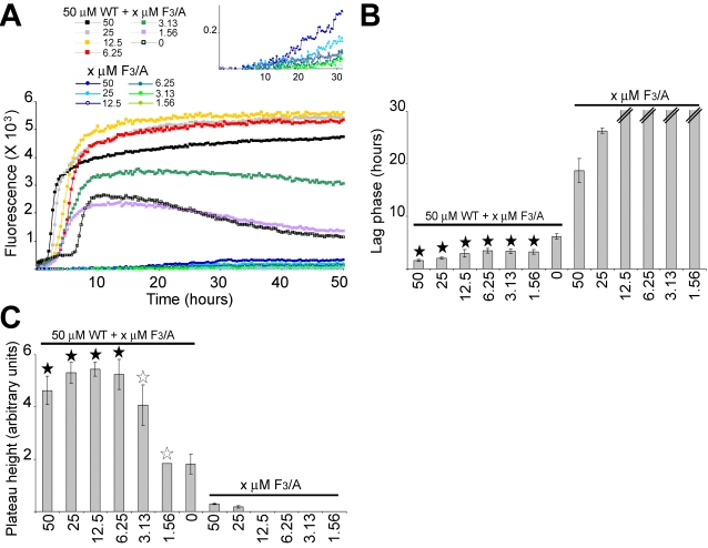
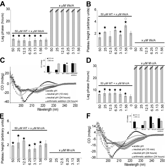
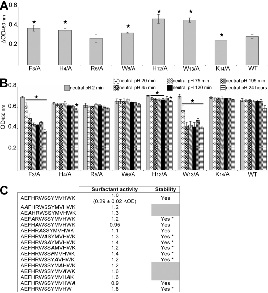
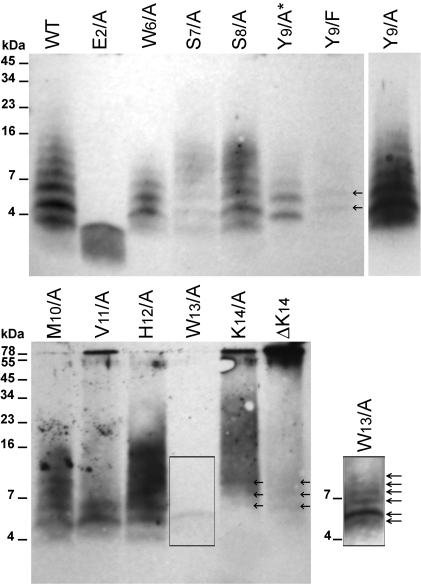
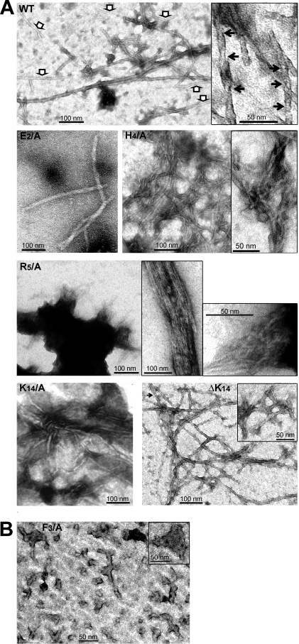
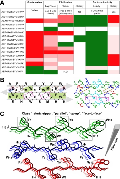
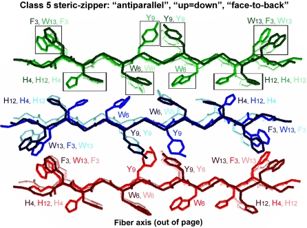
Polymerization into amyloid fibrils is a crucial step in the pathogenesis of neurodegenerative syndromes. Amyloid assembly is governed by properties of the sequence backbone and specific side-chain interactions, since fibrils from unrelated sequences possess similar structures and morphologies. Therefore, characterization of the structural determinants driving amyloid aggregation is of fundamental importance. We investigated the forces involved in the amyloid assembly of a model peptide derived from the oligomerization domain of acetylcholinesterase (AChE), AChE(586-599), through the effect of single point mutations on beta-sheet propensity, conformation, fibrilization, surfactant activity, oligomerization and fibril morphology. AChE(586-599) was chosen due to its fibrilization tractability and AChE involvement in Alzheimer's disease. The results revealed how specific regions and residues can control AChE(586-599) assembly. Hydrophobic and/or aromatic residues were crucial for maintaining a high beta-strand propensity, for the conformational transition to beta-sheet, and for the first stage of aggregation. We also demonstrated that positively charged side-chains might be involved in electrostatic interactions, which could control the transition to beta-sheet, the oligomerization and assembly stability. Further interactions were also found to participate in the assembly. We showed that some residues were important for AChE(586-599) surfactant activity and that amyloid assembly might preferentially occur at an air-water interface. Consistently with the experimental observations and assembly models for other amyloid systems, we propose a model for AChE(586-599) assembly in which a steric-zipper formed through specific interactions (hydrophobic, electrostatic, cation-pi, SH-aromatic, metal chelation and polar-polar) would maintain the beta-sheets together. We also propose that the stacking between the strands in the beta-sheets along the fiber axis could be stabilized through pi-pi interactions and metal chelation. The dissection of the specific molecular recognition driving AChE(586-599) amyloid assembly has provided further knowledge on such poorly understood and complicated process, which could be applied to protein folding and the targeting of amyloid diseases.


Conflict of interest statement

Competing Interests: Synaptica Ltd holds patents on the use of the AChE586-599 peptide and related peptides within T40 as potential biomarkers for neurodegenerative disease. The University of Oxford holds patents on the method of plate-based surface tension measurement.

Figures

Figure 1
Figure 1. Secondary structure propensity of AChE586-599 mutants as predicted by hidden β-propensity method (available at http://opal.umdnj.edu).
Propensities for helices (red squares), β-strands (blue squares) and random coil (green squares) are presented numerically using a 0-1 scale, with low values indicating zero to low propensity and high values indicating high propensity to near certainty. Hydrophobic residues are shown in green and aromatic residues in bold with a bigger font size.
Figure 2
Figure 2. Conformation of AChE586-599 and AChE586-599 mutants.
(A) Far UV spectra (250 to 190 nm) before and after pH neutralization (50 mM NaH2PO4, pH 7.2) of 4 AChE586-599 mutants (100 µM). These spectra are representatives of the different structures and different changes in structure observed for the wild-type and mutant peptides. (B) Conformation and changes in conformation after pH neutralization (50 mM NaH2PO4, pH 7.2) for AChE586-599 and all AChE586-599 mutants (100 µM). (C) Near UV spectra (320 to 240 nm) before and after pH neutralization (50 mM NaH2PO4, pH 7.2) of AChE586-599 and 4 mutants (100 µM). In all panels, the mutation within AChE586-599 is indicated in bold and italics.
Figure 3
Figure 3. Fibrilization properties of AChE586-599 and AChE586-599 mutants.
(A) 100 µM peptide was incubated with 165 µM ThT in PBS. Changes in ThT fluorescence were monitored (A, with the right panel showing scale-up of the left panel to visualize the rapid fibrilization of some peptides) with the lag phase of fibrilization (B, left panel) and plateau height (B, right panel) depicted. A black star signifies p<0.003 (B, left panel) and p<0.03 (B, right panel) when compared to the wild type peptide (WT). The peptides shown are representatives of the different fibrilization properties observed. (C) Fibrilization properties for AChE586-599 and all AChE586-599 mutants (100 µM). The properties are divided into 4 categories: ability to fibrilize, duration of lag phase, height of plateau and stability of the amyloid products (indicated by stability or decay of the ThT fluorescence after plateau). The mutation within AChE586-599 is indicated in bold and italics. The peptides that do not fibrilize and/or the peptides which amyloid products are not stable are indicated by grey boxes. The lag phase and plateau height for the mutant peptides are shown as fold ratio of AChE586-599 (e.g. ‘1’ represents equal value to AChE586-599 and ‘100’ for the lag phase represents 100 times longer than AChE586-599). ‘N.D.’ means ‘not detectable’.
Figure 4
Figure 4. Effect of ionic strength on the fibrilization properties of mutant peptide with unstable amyloid products.
Mutant peptide E2/A (50 µM) (A and B) or K14/A (100 µM) (C) were incubated with 165 µM ThT in 1.8 mM KH2PO4 and 10.1 mM NaH2PO4 with varying concentration of NaCl and KCl. The concentrations of NaCl and KCl were respectively: 0 and 0 (0 X), 42.8 mM and 0.84 mM (0.31 X), 85.6 mM and 1.7 mM (0.63 X), 136.9 mM and 2.7 mM (1 X), 171.1 mM and 3.4 mM (1.25 X), 350 mM and 6.7 mM (2.5 X), 700 mM and 13.5 mM (5 X), and 1.4 M and 27 mM (10 X). Changes in ThT fluorescence were monitored (A, with the right panel showing a scale-up of the left panel to visualize the rapid fibrilizations; and C). The lag phase of fibrilization (B, left panel) and plateau height (B, right panel; and C, right panel) are depicted. A black star signifies p<0.02 (B, left panel) and p<0.03 (B, right panel; C, right panel) when compared to 1 X NaCl and KCl.
Figure 5
Figure 5. Effect of the mutant peptide F3/A on AChE586-599 fibrilization.
Varying concentrations of the mutant F3/A were incubated with 165 µM ThT, with or without 50 µM AChE586-599. Changes in ThT fluorescence were monitored (A) with the lag phase of fibrilization (B) and the plateau height (C) depicted. The mutant peptide F3/A decreases the lag phase of AChE586-599 (A, with the inset showing a scale-up to visualize the fibrilization of the F3/A mutant on its own; and B), and increases the plateau height of AChE586-599 (A and C). A black star signifies p<0.03 (B) and p<0.05 (C) when compared to both 50 µM AChE586-599 and the equivalent concentration of the F3/A mutant. A white star signifies p<0.05 (C) when compared to the equivalent concentration of the F3/A mutant. The double bar in B indicates the absence of fibrilization (i.e. an indeterminably long lag phase).
Figure 6
Figure 6. Effect of the mutant peptides W6/A and M10/A on AChE586-599 fibrilization.
Varying concentrations of the mutant peptides were incubated with 165 µM ThT, with or without 50 µM AChE586-599. Changes in ThT fluorescence were measured and plotted as the lag phase of fibrilization (A and D) and the plateau height (B and E). The double bar in A and D indicates the absence of fibrilization (i.e.an indeterminably long lag phase). Far UV spectra (250 to 190 nm) before and after pH neutralization (50 mM NaH2PO4, pH 7.2) of 75 µM AChE586-599 with 75 µM mutant peptide (C and F). The insets show the mean residue ellipticity at 200 nm (random coil) and 215 nm (β-sheet) for 75 µM AChE586-599, 75 µM mutant peptide, 75 µM AChE586-599 with 75 µM mutant peptide, and the arithmetic addition of AChE586-599 to the mutant. (A and B) The mutant peptide W6/A does not affect either the lag phase (A, a black star signifies p<0.002 when compared to W6/A at the equivalent concentration) or the plateau height of AChE586-599 (B, a black star signifies p<0.045 when compared to W6/A at the equivalent concentration; a white star signifies p<0.05 when compared to AChE586-599). (C) The mutant peptide W6/A interacts with AChE586-599. A black star signifies p<0.014 when compared to the arithmetic addition of 75 µM W6/A mutant to 75 µM AChE586-599. (D and E) The mutant peptide M10/A does not affect either the lag phase (D, a black star signifies p<0.0007 when compared to M10/A at the equivalent concentration) or the plateau height of AChE586-599 (E, a black star signifies p<0.045 when compared to M10/A at the equivalent concentration). (F) The mutant peptide M10/A interacts with AChE586-599. A black star signifies p<0.01 when compared to the arithmetic addition of 75 µM M10/A mutant and 75 µM AChE586-599.
Figure 7
Figure 7. Surfactant properties of AChE586-599 and AChE586-599 mutants.
Surface tension was measured before and after neutralization (1M NaH2PO4, pH 7.2). (A) Representative surfactant activity of the AChE586-599 mutants (50 µM). ΔOD calculations were as described in Methods. A black star signifies p<0.035 when compared to AChE586-599. (B) Temporal pattern of the surfactant properties for AChE586-599 and AChE586-599 mutants. The peptides shown are representatives of the different surfactant properties observed. A black star signifies p<0.05 when compared to the same peptide after 2 min at neutral pH. (C) Surfactant properties for AChE586-599 and all AChE586-599 mutants (50 µM). The properties are divided into 2 categories: surfactant activity dependent on pH (depicted by subtracting the value at acidic pH to the value at neutral pH after 2 min) and stability of the surfactant activity (indicated by stability or decay of the OD signal). The mutation within AChE586-599 is indicated in bold and italics. The peptides with unstable surfactant activity are indicated by grey boxes. ‘*’ indicates peptides which activity remains stable over the time course, albeit one time point. The activity for the mutant peptides is shown as fold ratio of AChE586-599 activity (e.g. ‘1’ represents equal value to AChE586-599).
Figure 8
Figure 8. Formation of amyloid oligomers by AChE586-599 and AChE586-599 mutants.
Oligomers of AChE586-599 and AChE586-599 mutants (12 µM) were cross-linked by photo-induced cross-linking. Cross-linked products were resolved (16.5% Tris-Tricine SDS-PAGE), electro-blotted onto nitrocellulose and probed with Mab 105A (specific for AChE586-599 in β-sheet conformation). Marker proteins are indicated. Arrows indicate low abundance oligomeric species. Due to the strength of the signal for the oligomeric species, Y9/A was loaded at a third of the amount of the other peptides (Y9/A*). The signal resulting from loading equal amount to the other peptides can be seen on the individual lane on the right hand side of the top panel (Y9/A). On the right hand side of the bottom panel, an overexposure of the signal for W13/A shows multiple oligomeric species not seen at normal exposure.
Figure 9
Figure 9. T40/IDE digestion products form amyloid protofibrils.
Electron micrographs of negatively stained AChE586-599 and AChE586-599 mutants showing fibrils (A) and protofibrils (B). The white arrows in the left panel indicate thinner fibrils, and the black arrows indicate twists in the fibrils (A).
Figure 10
Figure 10. Structural model for AChE586-599 amyloid assembly based on conformation, fibrilization and surfactant properties of the wild type and mutant peptides.
(A) Summary of the conformation, fibrilization and surfactant properties of AChE586-599 and mutant peptides. Mutant peptides with similar properties to AChE586-599 (white boxes), with enhanced properties (green boxes), with diminished properties (red boxes, with low decrease indicated by light red and strong decrease by dark red). ‘N.D.’ indicates ‘non-detectable’. (B) Model of interactions between residues of AChE586-599 β-strands, which form a steric-zipper interface between the fibril forming β-sheets. The steric-zipper interface is shown as an antiparallel assembly of three AChE586-599 β-strands. AChE586-599 is represented at the primary amino acid sequence level (left panel) or at the carbon backbone structure level (right panel). On the left panel, hydrophobic interactions are represented as green shaded boxes, electrostatic interactions as blue and red shaded boxes, cation-π interactions as pink boxes and potential metal binding sites as brown boxes. The grey arrows indicate the direction of the strands. On the right panel, the chains are colored by residue type with hydrophobic residues (A, F, W, M and V) in green, negatively charged (E) in red, positively charged (H, R and K) in blue, and polar (S and Y) in cyan. (C) Model of quaternary interactions within β-sheets of AChE586-599 within a fibril. Each β-sheet is represented with only 3 copies of AChE586-599 for clarity: sheet 1 colored in different shades of green, sheet 2 in different shades of blue, and sheet 3 in different shades of red. The fibril is growing from the lighter to the darker color. On the carbon backbone structure, only the side chains of aromatic residues (F3, H4, W6, Y9, H12 and W13) are represented for clarity. The boxes highlight possible aromatic interactions (π-π) between strands within a β-sheet. Within a β-sheet, AChE586-599 strands are stacking in a parallel arrangement. Within AChE586-599 fibril, the β-sheets are antiparallel, have the same sides facing each other (‘face-to-face’) and the orientation of the sheet edges facing up (‘up-up’). According to the nomenclature of Sawaya et al., this type of arrangement and orientation corresponds to a class 1 steric-zipper .
Figure 11
Figure 11. Model of quaternary interactions within β-sheets of AChE586-599 within a fibril of class 5.
Each β-sheet is represented with only 3 copies of AChE586-599 for clarity: sheet 1 colored in different shades of green, sheet 2 in different shades of blue, and sheet 3 in different shades of red. On the carbon backbone structure, only the side chains of aromatic residues (F3, H4, W6, Y9, H12 and W13) are represented for clarity. The boxes highlight possible aromatic interactions (π-π) between strands within a β-sheet. Within a β-sheet, AChE586-599 strands are stacking in an antiparallel arrangement. Within AChE586-599 fibril, the β-sheets are antiparallel, have the same sides facing each other (‘face-to-face’), however the orientation of the strand edges within a sheet alternate between up and down (‘up = down’). According to the nomenclature of Sawaya et al., this type of arrangement and orientation corresponds to a class 5 steric-zipper .

References

    1. Harper JD, Lansbury PT., Jr Models of amyloid seeding in Alzheimer's disease and scrapie: mechanistic truths and physiological consequences of the time-dependent solubility of amyloid proteins. Annu Rev Biochem. 1997;66:385–407. - PubMed
    1. Westermark P. Aspects on human amyloid forms and their fibril polypeptides. Febs J. 2005;272:5942–5949. - PubMed
    1. Klunk WE, Pettegrew JW, Abraham DJ. Quantitative evaluation of congo red binding to amyloid-like proteins with a beta-pleated sheet conformation. J Histochem Cytochem. 1989;37:1273–1281. - PubMed
    1. LeVine H., 3rd Thioflavine T interaction with synthetic Alzheimer's disease beta-amyloid peptides: detection of amyloid aggregation in solution. Protein Sci. 1993;2:404–410. - PMC - PubMed
    1. Rochet JC, Lansbury PT., Jr Amyloid fibrillogenesis: themes and variations. Curr Opin Struct Biol. 2000;10:60–68. - PubMed

Publication types

MeSH terms