. 2008 Mar 19;3(3):e1852.
doi: 10.1371/journal.pone.0001852.

Hexokinase II detachment from mitochondria triggers apoptosis through the permeability transition pore independent of voltage-dependent anion channels

Affiliations

Hexokinase II detachment from mitochondria triggers apoptosis through the permeability transition pore independent of voltage-dependent anion channels

Federica Chiara et al. PLoS One. .

Abstract

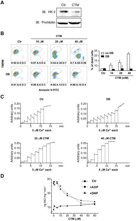
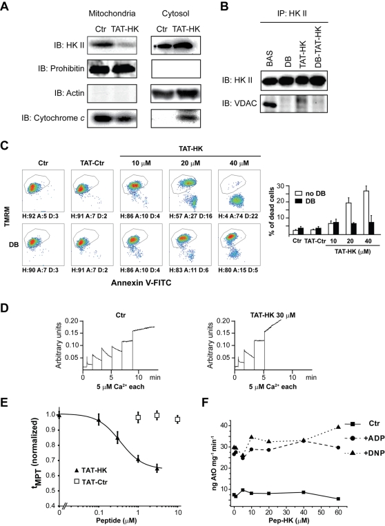
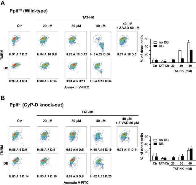
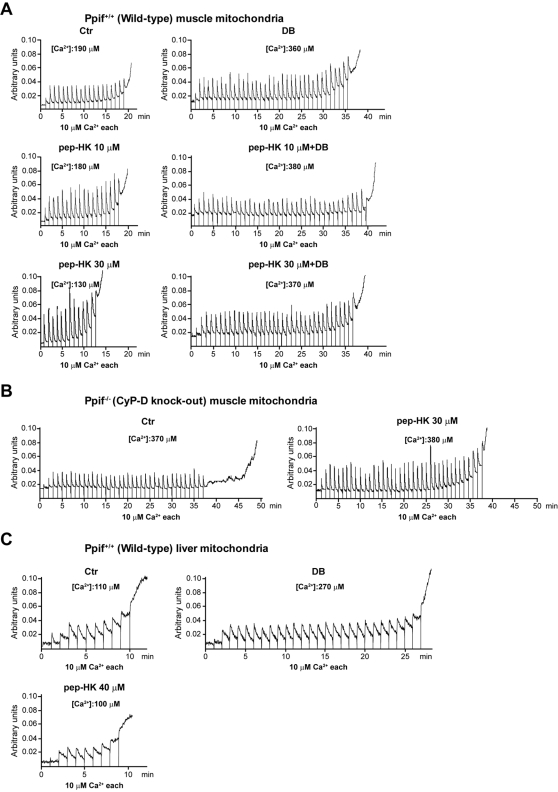
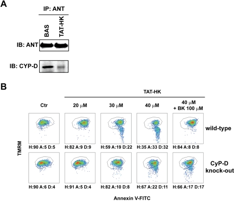
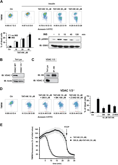
Type II hexokinase is overexpressed in most neoplastic cells, and it mainly localizes on the outer mitochondrial membrane. Hexokinase II dissociation from mitochondria triggers apoptosis. The prevailing model postulates that hexokinase II release from its mitochondrial interactor, the voltage-dependent anion channel, prompts outer mitochondrial membrane permeabilization and the ensuing release of apoptogenic proteins, and that these events are inhibited by growth factor signalling. Here we show that a hexokinase II N-terminal peptide selectively detaches hexokinase II from mitochondria and activates apoptosis. These events are abrogated by inhibiting two established permeability transition pore modulators, the adenine nucleotide translocator or cyclophilin D, or in cyclophilin D knock-out cells. Conversely, insulin stimulation or genetic ablation of the voltage-dependent anion channel do not affect cell death induction by the hexokinase II peptide. Therefore, hexokinase II detachment from mitochondria transduces a permeability transition pore opening signal that results in cell death and does not require the voltage-dependent anion channel. These findings have profound implications for our understanding of the pathways of outer mitochondrial membrane permeabilization and their inactivation in tumors.


Conflict of interest statement

Competing Interests: The authors have declared that no competing interests exist.

Figures

Figure 1
Figure 1. Effects of clotrimazole on cells and mitochondria.
(A) HK II detachment from mitochondria following a 1 hour treatment with clotrimazole (CTM, 20 µM) was assessed by Western immunoblot on a mitochondrial fraction of HeLa cells. The blot was also probed with prohibitin as a mitochondrial marker and with actin as a cytosolic marker (negative, not shown), to check for fraction purity. (B) Output of multiparametric FACS analysis show apoptosis induction of HeLa cells exposed for 2 hours to several concentrations of CTM with or without pre-incubation with Debio 025 (DB, 8 µM). Healthy cells (H) are delimited by the quadrant. Apoptotic cells (A) display mitochondrial depolarization (reduced TMRM staining) and/or expose phosphatidylserine on their surface (increased Annexin V-FITC staining). Propidium iodide-positive, i.e. dead cells (D), were evaluated on a PI vs TMRM diagram (not shown); these cells were excluded from the reported plots and shown as histograms. Numbers are percentages. (C) PTP opening of digitonized HeLa cells measured with the Ca2+ retention capacity assay. Calcium Green-5N fluorescence is reported as arbitrary units on the y axis. As the probe does not permeate mitochondria, Ca2+ uptake into the organelles is displayed by a rapid decrease of the fluorescence spike. Pore inducers or inhibitors reduce or increase, respectively, the threshold Ca2+ concentration required to trigger the permeability transition, i.e. the number of spikes before a sudden and marked fluorescence increase occurs. Experiments were started by the addition of digitonized cells (not shown) followed by pulses of the indicated concentrations of Ca2+. Where indicated, CTM was added to cells 45 minutes before permeabilization with digitonin. Note that CTM reduced the rate of Ca2+ uptake into mitochondria. In the upper right plot DB was added before mitochondria to test the PTP-dependence of Ca2+ release. (D) Respiration assay performed on mitochondria isolated from mouse liver. The graph shows the rate of oxygen consumption (ng AtO stands for ngatoms of oxygen) vs. different concentrations of CTM. The rate of respiration is displayed in basal conditions (▪), after ADP administration (•) or after dinitrophenol administration (▴). All reported results in the Figure are representative of at least four experiments.
Figure 2
Figure 2. Selective detachment of HK II from mitochondria induces PTP opening and cell death.
(A) HK II detachment from mitochondria of HeLa cells following a 1-hour treatment with TAT-HK (20 µM). Western immunoblots of a mitochondrial and of a cytosolic fraction show a redistribution of both HK II and cytochrome c into the cytosol. Blots were also probed with prohibitin as a mitochondrial marker and with actin as a cytosolic marker to check for fraction purity. (B) HK II immunoprecipitation in HeLa cells kept in control conditions or treated for 1 hour with Debio 025 (DB, 8 µM), TAT-HK (20 µM), or both. Co-immunoprecipitation of VDAC is shown. (C) Output of multiparametric FACS analyses show apoptosis induction of HeLa cells exposed for 2 hours to a control peptide linked to the TAT sequence (TAT-Ctr, 40 µM) or to the reported concentrations of TAT-HK with or without pre-incubation with Debio 025 (DB, 8 µM). Diagrams and percentages of the different cell populations are as in Fig. 1B. (D) PTP opening of digitonized HeLa cells measured with the Ca2+ retention capacity assay. Experiments were started by the addition of cells (not shown) followed by pulses of the indicated concentrations of Ca2+; where indicated, TAT-HK (30 µM) was added to cells 45 minutes before permeabilization with digitonin. TAT-Ctr (40 µM) did not change the number of Ca2+ pulses (not shown). Calcium Green-5N fluorescence is reported as arbitrary units on the y axis. (E) MPT susceptibility to ROS (tMPT) in intact cardiac myocytes is significantly enhanced by TAT-HK while the negative control peptide TAT-Ctr has no effect. tMPT measurements were performed after 1 hr treatment with peptides. Figure represents four independent experiments with 50–60 cells per groups examined. (F) Respiration assay performed on mouse liver mitochondria. The rate of oxygen consumption (ng AtO stands for ngatoms of oxygen) is plotted vs. different concentrations of the HK peptide. The rate of respiration is displayed in basal conditions (▪), after ADP administration (•) or after dinitrophenol administration (▴). All reported measures in the Figure are representative of at least four experiments.
Figure 3
Figure 3. CyP-D modulates apoptosis triggered by detachment of HK II from mitochondria.
(A, B) Output of multiparametric FACS analyses show apoptosis induction in fibroblasts obtained either from wild-type mice (A) or from mice in which the Ppif gene encoding for CyP-D was ablated (B). Cells were exposed for 2 hours to the reported concentrations of TAT-HK with or without pre-incubation with Debio 025 (DB, 8 µM) or with a pan-caspase inhibitor (Z.VAD-fmk, 50 µM). Diagrams and percentages of the different cell populations are indicated as in Fig. 1B. All reported measures in the Figure are representative of at least four experiments.
Figure 4
Figure 4. CyP-D modulates PTP opening triggered by detachment of HK II from mitochondria.
CRC experiments were performed on mitochondria isolated from muscle of either wild-type (A) or CyP-D knock-out mice (B), or from liver of wild-type animals (C). Assays were started by the addition of mitochondria followed by pulses of the indicated concentrations of Ca2+; where indicated, the reported concentrations of the HK peptide lacking the TAT sequence and/or Debio 025 (DB, 8 µM) were added to mitochondria 5 minutes before recordings. Calcium Green-5N fluorescence is reported as arbitrary units on the y axis. Total Ca2+ loading required to open the pore is reported for each condition, and all reported results are representative of at least four experiments. Treatment of mitochondria with the control peptide (40 µM) or with a HK peptide harbouring the TAT sequence did not change the number of Ca2+ pulses (not shown). All reported measures in the Figure are representative of at least four experiments.
Figure 5
Figure 5. ANT modulates apoptosis triggered by detachment of HK II from mitochondria.
(A) Western blot of ANT immunoprecipitation. Co-immunoprecipitation of CyP-D in wild-type mouse fibroblasts is shown either in control conditions or after a 1 hour treatment with TAT-HK. (B) Output of multiparametric FACS analyses show apoptosis induction in fibroblasts obtained either from wild-type mice (upper row) or from CyP-D knock-out animals (lower row). Cells were exposed for 2 hours to the stated concentrations of TAT-HK with or without a 3 hour pre-incubation with bongkrekate (BK, 100 µM). Diagrams and percentages of the different cell populations are indicated as in Fig. 1B. All reported measures in the Figure are representative of at least four experiments.
Figure 6
Figure 6. Neither insulin nor VDAC modulate apoptosis triggered by detachment of HK II from mitochondria.
(A) After 10 minutes of insulin (5 µg/ml) stimulation, HeLa cells were exposed for 2 hours to the reported concentrations of TAT-HK and apoptosis assayed as in Figure 1B. Western immunoblot shows GSK3β inhibition (i.e. Ser9 phosphorylation) by insulin. (B) HK II and VDAC expression in MEF obtained from either wild-type or VDAC 1/3 knock-out mice were assessed by Western immunoblot. VDAC expression was checked with an anti-VDAC1antibody, protein loading with an anti-actin antibody. (C) Immunoprecipitation of HK II and rehybridization with an anti VDAC2 in VDAC1/3 knock-out MEFs. (D) Output of multiparametric FACS analyses show apoptosis induction in VDAC1/3 knock-out MEFs. Cells were exposed for 1 hour to the stated concentrations of TAT-HK with or without pre-incubation with Debio 025 (DB, 8 µM), bongkrekate (BK, 100 µM) or Z.VAD-fmk (50 µM). Diagrams and percentages of the different cell populations are indicated as in Fig. 1B. (E) Mitochondrial depolarization after cell treatment with TAT-HK (10 µM) with or without pre-incubation with Debio 025 (8 µM), as assessed by decreased TMRM staining by epifluorescence microscopy. All reported measurements are representative of at least four experiments.

References

    1. Wilson JE. Isozymes of mammalian hexokinase: structure, subcellular localization and metabolic function. J Exp Biol. 2003;206:2049–2057. - PubMed
    1. Mathupala SP, Ko YH, Pedersen PL. Hexokinase II: cancer's double-edged sword acting as both facilitator and gatekeeper of malignancy when bound to mitochondria. Oncogene. 2006;25:4777–4786. - PMC - PubMed
    1. Warburg O. On the origin of cancer cells. Science. 1956;123:309–314. - PubMed
    1. Majewski N, Nogueira V, Bhaskar P, Coy PE, Skeen JE, et al. Hexokinase-mitochondria interaction mediated by Akt is required to inhibit apoptosis in the presence or absence of Bax and Bak. Mol Cell. 2004;16:819–830. - PubMed
    1. Rathmell JC, Fox CJ, Plas DR, Hammerman PS, Cinalli RM, et al. Akt-directed glucose metabolism can prevent Bax conformation change and promote growth factor-independent survival. Mol Cell Biol. 2003;23:7315–7328. - PMC - PubMed

Publication types