Detection of human cytomegalovirus in different histological types of gliomas
- PMID: 18351367
- PMCID: PMC3001277
- DOI: 10.1007/s00401-008-0359-1
Detection of human cytomegalovirus in different histological types of gliomas
Abstract
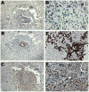
The association between human cytomegalovirus (HCMV) infection and glioblastoma has been a source of debate in recent years because of conflicting laboratory reports concerning the presence of the virus in glioma tissue. HCMV is a ubiquitous herpesvirus that exhibits tropism for glial cells and has been shown to transform cells in vitro. Using sensitive immunohistochemical and in situ hybridization methods in 50 glioma samples, we detected HCMV antigen and DNA in 21/21 cases of glioblastoma, 9/12 cases of anaplastic gliomas and 14/17 cases of low-grade gliomas. Reactivity against the HCMV IE1 antigen (72 kDa) exhibited histology-specific patterns with more nuclear staining for anaplastic and low-grade gliomas, while GBMs showed nuclear and cytoplasmic staining that likely occurs with latent infection. Using IHC, the number of HCMV-positive cells in GBMs was 79% compared to 48% in lower grade tumors. Non-tumor areas of the tissue contained only four and 1% of HCMV-positive cells for GBMs and lower grade tumors, respectively. Hybridization to HCMV DNA in infected cells corresponded to patterns of immunoreactivity. Our findings support previous reports of the presence of HCMV infection in glioma tissues and advocate optimization of laboratory methods for the detection of active HCMV infections. This will allow for detection of low-level latent infections that may play an important role in the initiation and/or promotion of malignant gliomas.
Figures


References
-
- Albrecht T, Boldogh I, Fons M, Lee CH, AbuBakar S, Russell JM, Au WW. Cell-activation responses to cytomegalovirus infection relationship to the phasing of CMV replication and to the induction of cellular damage. Subcell Biochem. 1989;15:157–202. - PubMed
-
- Albrecht T, Fons MP, Deng CZ, Boldogh I. Increased frequency of specific locus mutation following human cytomegalovirus infection. Virology. 1997;230:48–61. - PubMed
-
- Albrecht T, Deng CZ, Abdel-Rahman SZ, Fons M, Cinciripini P, El Zein RA. Differential mutagen sensitivity of peripheral blood lymphocytes from smokers and nonsmokers: effect of human cytomegalovirus infection. Environ Mol Mutagen. 2004;43:169–178. - PubMed
-
- Arbustini E, Morbini P, Grasso M, Diegoli M, Fasani R, Porcu E, Banchieri N, Perfetti V, Pederzolli C, Grossi P, Dalla Gasperina D, Martinelli L, Paulli M, Ernst M, Plachter B, Vigano M, Solcia E. Human cytomegalovirus early infection, acute rejection, and major histocompatibility class II expression in transplanted lung. Molecular, immunocytochemical, and histopathologic investigations. Transplantation. 1996;61:418–427. - PubMed
-
- Bagasra O, Hansen JA, editors. In-Situ PCR Techniques. Wiley, John & Sons; New York: 2006.
Publication types
MeSH terms
Substances
Grants and funding
LinkOut - more resources
Full Text Sources
Other Literature Sources
Medical

