Case Reports
. 2008 Mar;1(3):247-54.
doi: 10.3171/PED/2008/1/3/247.

Identification of distinct and overlapping cortical areas for bilingual naming and reading using cortical stimulation. Case report

Affiliations
Case Reports

Identification of distinct and overlapping cortical areas for bilingual naming and reading using cortical stimulation. Case report

Sandra Serafini et al. J Neurosurg Pediatr. 2008 Mar.

Abstract

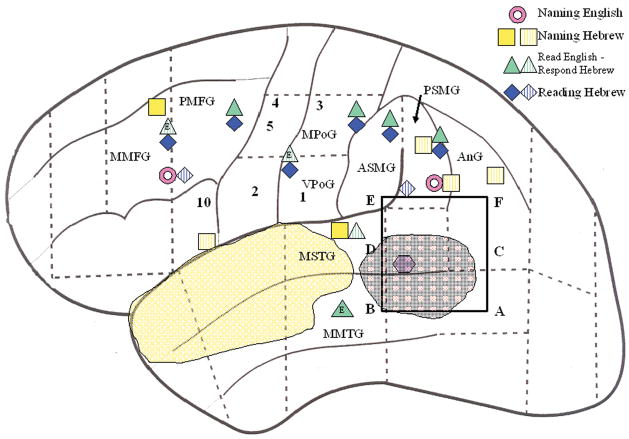
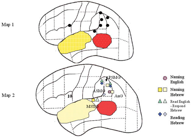
A bilingual pediatric patient who underwent tumor resection was mapped extraoperatively using cortical stimulation to preserve English and Hebrew languages. The authors mapped both languages by using 4 tasks: 1) English visual naming, 2) Hebrew visual naming, 3) read English/respond Hebrew, and 4) Hebrew reading. Essential cortical sites for primary and secondary languages were compared, photographically recorded, and plotted onto a schematic brain of the patient. Three types of sites were found in this patient: 1) multiuse sites (multiple tasks, both languages) in frontal, temporal, and parietal areas; 2) single-task sites (1 task, both languages) in postcentral and parietal areas; and 3) single-use sites (1 task, 1 language) in frontal, temporal, and parietal areas. These results lend support to the concept that bilingual patients can have distinct cortical representations of each language and of different language tasks, in addition to overlapping or shared sites that support both languages and multiple tasks.


Figures

Figure 1
Figure 1
A. T-2 weighted spin-echo axial anatomical image of resection cavity and recurrent tumor (GE Signa LX 1.5T, AC-PC slice orientation, TR/TE/Flip angle = 3000ms/84ms/90°, matrix = 256 × 256, FOV = 24cm, voxels = 0.92 × 0.92 × 5.0mm). The image shows the resection of portions of left superior and middle temporal gyrus (yellow outline) and recurrent tumor posterior to the cavity (red outline). B. Schematic showing approximate area of the resection area (yellow), recurrent tumor (red), and English visual naming sites (black).
Figure 1
Figure 1
A. T-2 weighted spin-echo axial anatomical image of resection cavity and recurrent tumor (GE Signa LX 1.5T, AC-PC slice orientation, TR/TE/Flip angle = 3000ms/84ms/90°, matrix = 256 × 256, FOV = 24cm, voxels = 0.92 × 0.92 × 5.0mm). The image shows the resection of portions of left superior and middle temporal gyrus (yellow outline) and recurrent tumor posterior to the cavity (red outline). B. Schematic showing approximate area of the resection area (yellow), recurrent tumor (red), and English visual naming sites (black).
Figure 2
Figure 2
Schematic of grid placement. First resection area shown in yellow, recurrent tumor in red with visible lesion in purple. Boxed electrodes, rows 1–3 show where a focal seizure was induced with 6mA stimulation; naming and reading in both languages remained intact. (contacts 1–4, 9–12, 17–20).
Figure 3
Figure 3
A) Tagged sites for visual naming, reading, and read/respond in English and Hebrew. Letters A-F show stimulation-induced seizure activity. 1=Tongue (sensory), 2=Mouth (motor), 3=Hand (sensory), 4=Thumb (motor), 5=Jaw movement, 10=Speech arrest (counting). One-centimeter measurement tag reference. B) Schematic map of English Naming (pink donut), Hebrew Naming (yellow squares), Read English/Respond Hebrew (green triangles; E = English reading error only), and Reading Hebrew (blue diamonds). Striped symbols represent p-values between 0.05 and 0.2. MFG = Middle Frontal Gyrus, PoG = Post Central Gyrus, SMG = Supramarginal Gyrus, AnG = Angular Gyrus, STG = Superior Temporal Gyrus, MTG = Middle Temporal Gyrus. Letters preceding these abbreviations refer to Posterior (P), Middle (M), Ventral (V).
Figure 3
Figure 3
A) Tagged sites for visual naming, reading, and read/respond in English and Hebrew. Letters A-F show stimulation-induced seizure activity. 1=Tongue (sensory), 2=Mouth (motor), 3=Hand (sensory), 4=Thumb (motor), 5=Jaw movement, 10=Speech arrest (counting). One-centimeter measurement tag reference. B) Schematic map of English Naming (pink donut), Hebrew Naming (yellow squares), Read English/Respond Hebrew (green triangles; E = English reading error only), and Reading Hebrew (blue diamonds). Striped symbols represent p-values between 0.05 and 0.2. MFG = Middle Frontal Gyrus, PoG = Post Central Gyrus, SMG = Supramarginal Gyrus, AnG = Angular Gyrus, STG = Superior Temporal Gyrus, MTG = Middle Temporal Gyrus. Letters preceding these abbreviations refer to Posterior (P), Middle (M), Ventral (V).
Figure 4
Figure 4
Map 1: English visual naming sites (black dots) from the first resection. Map 2: Proximate sites from all tasks in second resection. 10=Speech arrest (counting), SMG = Supramarginal Gyrus, AnG = Angular Gyrus, STG = Superior Temporal Gyrus. Letters preceding these abbreviations refer to Posterior (P), Middle (M), Anterior (A).

References

    1. Bates E, Reilly J, Wulfeck B, Dronkers N, Opie M, Fenson J, Kriz S, Jeffries R, Miller L, Herbst K. Differential effects of unilateral lesions on language production in children and adults. Brain and Language. 2001;79:223–265. - PubMed
    1. Belin P, Van Eeckhout P, Zilbovicious M, Remy P, Francois C, Guillaume S, Chain F, Rancurel G, Samson Y. Recovery from nonfluent aphasia after melodic intonation therapy: A PET Study. Neurology. 1996;47:1504–1511. - PubMed
    1. Bookheimer S, Zeffiro T, Blaxton T, Gaillard W, Theodore W. Regional cerebral blood flow changes during object naming and word reading. Human Brain Mapping. 1995;3:93–106.
    1. Cao Y, Vikingstad EM, George KP, Johnson AF, Welch KMA. Cortical language activation in stroke patients recovering from aphasia with functional MRI. Stroke. 1999;30:2331–2340. - PubMed
    1. Chee M, Tan E, Thiel T. Mandarin and English single word processing studied with functional magnetic resonance imaging. The Journal of Neuroscience. 1999a;19:3050–3056. - PMC - PubMed

Publication types

LinkOut - more resources