

Distribution of SIBLING proteins in the organic and inorganic phases of rat dentin and bone
- PMID: 18353003
- PMCID: PMC2666982
- DOI: 10.1111/j.1600-0722.2008.00522.x
Distribution of SIBLING proteins in the organic and inorganic phases of rat dentin and bone
Abstract
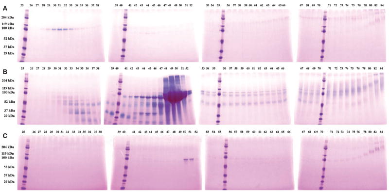
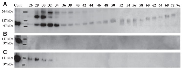
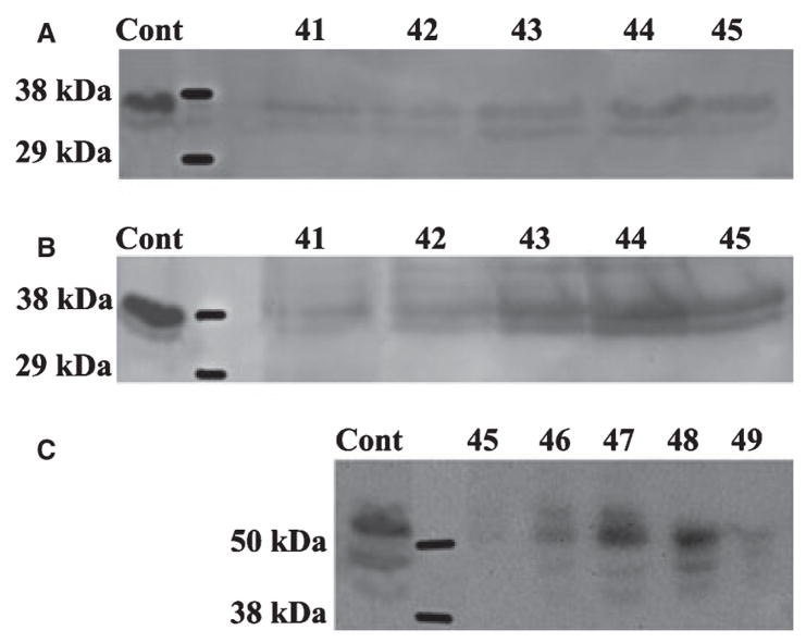
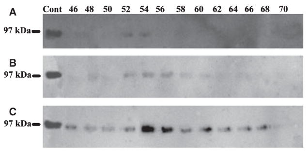
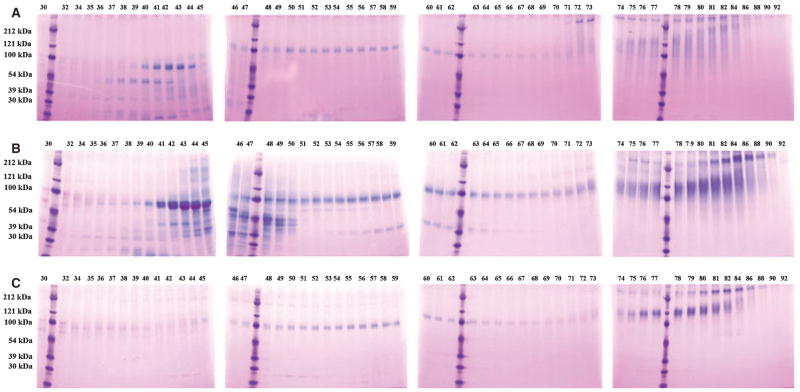
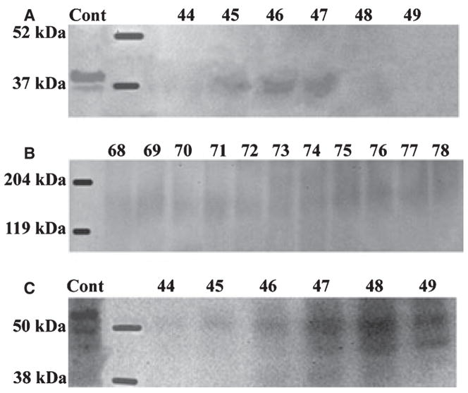
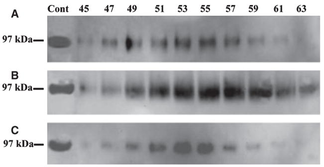
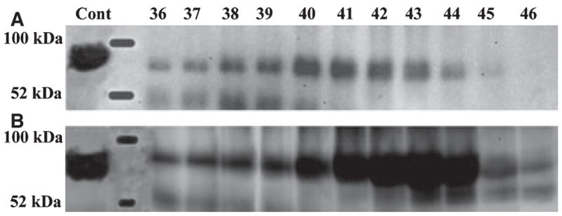
The SIBLING protein family is a group of non-collagenous proteins (NCPs) that includes dentin sialophosphoprotein (DSPP), dentin matrix protein 1 (DMP1), bone sialoprotein (BSP), and osteopontin (OPN). In the present study, we compared these four proteins in different phases of rat dentin and bone. First, we extracted NCPs in the unmineralized matrices and cellular compartments using guanidium-HCl (G1). Second, we extracted NCPs closely associated with hydroxyapatite using an EDTA solution (E). Last, we extracted the remaining NCPs again with guanidium-HCl (G2). Each fraction of Q-Sepharose ion-exchange chromatography was analyzed using sodium dodecyl sulfate-polyacrylamide gel electrophoresis (SDS-PAGE), Stains-All stain, and with western immunoblotting. In dentin, the NH(2)-terminal fragment of DSPP and its proteoglycan form were primarily present in the G1 extract, whereas the COOH-terminal fragment of DSPP was present exclusively in the E extract. The processed NH(2)-terminal fragment of DMP1 was present in G1 and E extracts, whereas the COOH-terminal fragment of DMP1 existed mainly in the E extract. Bone sialoprotein was present in all three extracts of dentin and bone, whereas OPN was present only in the G1 and E extracts of bone. The difference in the distribution of the SIBLING proteins between organic and inorganic phases supports the belief that these molecular species play different roles in dentinogenesis and osteogenesis.
Figures
Similar articles
-
Distribution of small integrin-binding ligand, N-linked glycoproteins (SIBLING) in the condylar cartilage of rat mandible.Int J Oral Maxillofac Surg. 2010 Mar;39(3):272-81. doi: 10.1016/j.ijom.2009.12.017. Epub 2010 Jan 25. Int J Oral Maxillofac Surg. 2010. PMID: 20097540 Free PMC article.
-
Expression and distribution of SIBLING proteins in the predentin/dentin and mandible of hyp mice.Oral Dis. 2010 Jul;16(5):453-64. doi: 10.1111/j.1601-0825.2010.01656.x. Epub 2010 Mar 9. Oral Dis. 2010. PMID: 20233318 Free PMC article.
-
Isolation of SIBLING Proteins from Bone and Dentin Matrices.Methods Mol Biol. 2019;1922:211-218. doi: 10.1007/978-1-4939-9012-2_21. Methods Mol Biol. 2019. PMID: 30838579
-
Post-translational modifications of sibling proteins and their roles in osteogenesis and dentinogenesis.Crit Rev Oral Biol Med. 2004 Jun 4;15(3):126-36. doi: 10.1177/154411130401500302. Crit Rev Oral Biol Med. 2004. PMID: 15187031 Review.
-
Hypophosphatemic osteosclerosis, hyperostosis, and enthesopathy associated with novel homozygous mutations of DMP1 encoding dentin matrix protein 1 and SPP1 encoding osteopontin: The first digenic SIBLING protein osteopathy?Bone. 2020 Mar;132:115190. doi: 10.1016/j.bone.2019.115190. Epub 2019 Dec 13. Bone. 2020. PMID: 31843680 Free PMC article. Review.
Cited by
-
Adhesion of MC3T3-E1 cells bound to dentin phosphoprotein specifically bound to collagen type I.J Biomed Mater Res A. 2012 Sep;100(9):2492-8. doi: 10.1002/jbm.a.34159. Epub 2012 May 21. J Biomed Mater Res A. 2012. PMID: 22615197 Free PMC article.
-
Probing the influence of SIBLING proteins on collagen-I fibrillogenesis and denaturation.Connect Tissue Res. 2018 May;59(3):274-286. doi: 10.1080/03008207.2017.1379514. Epub 2017 Oct 4. Connect Tissue Res. 2018. PMID: 28910556 Free PMC article.
-
Differential gene expression in the perichondrium and cartilage of the neonatal mouse temporomandibular joint.Orthod Craniofac Res. 2009 Aug;12(3):168-77. doi: 10.1111/j.1601-6343.2009.01450.x. Orthod Craniofac Res. 2009. PMID: 19627518 Free PMC article.
-
Tooth dentin defects reflect genetic disorders affecting bone mineralization.Bone. 2012 Apr;50(4):989-97. doi: 10.1016/j.bone.2012.01.010. Epub 2012 Jan 26. Bone. 2012. PMID: 22296718 Free PMC article. Review.
-
Gene evolution and functions of extracellular matrix proteins in teeth.Orthod Waves. 2013 Mar 1;72(1):1-10. doi: 10.1016/j.odw.2013.01.040. Epub 2013 Feb 23. Orthod Waves. 2013. PMID: 23539364 Free PMC article.
References
-
- Xiao S, Yu C, Chou X, Yuan W, Wang Y, Bu L, Fu G, Qian M, Yang J, Shi Y, Hu L, Han B, Wang Z, Huang W, Liu J, Chen Z, Zhao G, Kong X. Dentinogenesis imperfecta 1 with or without progressive hearing loss is associated with distinct mutations in DSPP. Nat Genet. 2001;27:201–204. - PubMed
-
- Zhang X, Zhao J, Li C, Gao S, Qiu C, Liu P, Wu G, Qiang B, Lo WH, Shen Y. DSPP mutation in dentinogenesis imperfecta Shields type II. Nat Genet. 2001;27:151–152. - PubMed
-
- Dong J, Gu T, Jeffords L, Macdougall M. Dentin phosphoprotein compound mutation in dentin sialophosphoprotein causes dentinogenesis imperfecta type III. Am J Med Genet A. 2005;132:305–309. - PubMed
-
- Sreenath T, Thyagarajan T, Hall B, Longenecker G, D’souza R, Hong S, Wright JT, Macdougall M, Sauk J, Kulkarni AB. Dentin sialophosphoprotein knockout mouse teeth display widened predentin zone and develop defective dentin mineralization similar to human dentinogenesis imperfecta type III. J Biol Chem. 2003;278:24874–24880. - PubMed
-
- Ye L, Macdougall M, Zhang S, Xie Y, Zhang J, Li Z, Lu Y, Mishina Y, Feng JQ. Deletion of dentin matrix protein-1 leads to a partial failure of maturation of predentin into dentin, hypomineralization, and expanded cavities of pulp and root canal during postnatal tooth development. J Biol Chem. 2004;279:19141–19148. - PubMed
Publication types
MeSH terms
Substances
Grants and funding
LinkOut - more resources
Full Text Sources
Research Materials
Miscellaneous
