Review
. 2008 Feb;21(1):39-55.
doi: 10.1111/j.1755-148X.2007.00427.x.

Cancer stem cells and human malignant melanoma

Affiliations
Review

Cancer stem cells and human malignant melanoma

Tobias Schatton et al. Pigment Cell Melanoma Res. 2008 Feb.

Abstract

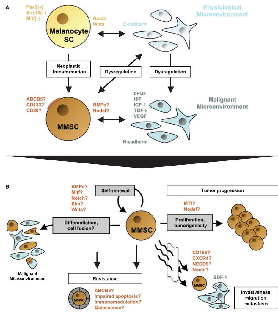
Cancer stem cells (CSC) have been identified in hematological malignancies and several solid cancers. Similar to physiological stem cells, CSC are capable of self-renewal and differentiation and have the potential for indefinite proliferation, a function through which they may cause tumor growth. Although conventional anti-cancer treatments might eradicate most malignant cells in a tumor, they are potentially ineffective against chemoresistant CSC, which may ultimately be responsible for recurrence and progression. Human malignant melanoma is a highly aggressive and drug-resistant cancer. Detection of tumor heterogeneity, undifferentiated molecular signatures, and increased tumorigenicity of melanoma subsets with embryonic-like differentiation plasticity strongly suggest the presence and involvement of malignant melanoma stem cells (MMSC) in the initiation and propagation of this malignancy. Here, we review these findings in the context of functional properties ascribed to melanocyte stem cells and CSC in other cancers. We discuss the association of deregulated signaling pathways, genomic instability, and vasculogenic mimicry phenomena observed in melanoma subpopulations in light of the CSC concept. We propose that a subset of MMSC may be responsible for melanoma therapy-resistance, tumor invasiveness, and neoplastic progression and that targeted abrogation of a MMSC compartment could therefore ultimately lead to stable remissions and perhaps cures of metastatic melanoma.


Figures

Figure 1
Figure 1
Schematic model of a potential relationship between melanocyte stem cells (Melanocyte SC) and putative malignant melanoma stem cells (MMSC) in the context of distinct physiological or malignant microenvironments, and overview of defining features and functional properties ascribed to putative MMSC. (A) Physiological melanocyte stem cells are characterized by a Pax3+, Sox10 and Mitf phenotype, which promotes immaturity and quiescence. The balance between melanocyte SC self-renewal and differentiation, quiescence and proliferation in the hair follicle is further regulated by Notch and/or Wnt signaling molecules. It is thought that the homeostasis of these lineage-restricted SC is maintained through interactions with the physiological microenvironment, mainly composed of E-cadherin-expressing keratinocytes. MMSC might arise by cumulative oncogenic mutations and/or loss of tumor suppressors from long-lived melanocyte stem cells, which already contain the machinery for indefinite self-renewal. Surface markers that have been associated with melanoma stem-like cells include ABCB5, CD133, or CD20. Moreover, morphogens such as Nodal or BMP4 may be secreted by putative MMSC populations. In addition to the neoplastic transformation of the melanocyte stem cell itself, distinct changes in the microenvironment may facilitate the formation of MMSC and subsequent MMSC-driven melanomagenesis. A dysregulated, malignant microenvironment described for melanocytic tumors is characterized by the production of large quantities of proliferative growth factors, including bFGF, HIF, IGF-1, TGF-β, and VEGF. These paracrine mediators, produced mainly by N-cadherin-expressing endothelial cells or surrounding fibroblasts, may drive MMSC proliferation and hence melanoma progression. On the other hand, MMSC might actively alter their surrounding microenvironment through secreted factors (i.e., Nodal or BMP), thus sustaining a niche hospitable to tumor initiation and growth. (B) Defining characteristics of putative MMSC populations and their potential involvement in melanoma initiation, progression, and resistance. Analogous to the cancer stem cell (CSC) definition, MMSC should exhibit (1) increased in vivo tumorigenicity, (2) in vivo self-renewal capacity, and (3) the competence to differentiate into phenotypically diverse populations, including non-tumorigenic cancer cells present in the original tumor. Regulatory signaling networks operative in physiological stem cells that have been shown to be dysregulated in malignant melanoma, such as BMP, Mitf, Notch, Shh, or Wnts, may likewise modulate the balance between self-renewal and differentiation of MMSC. Molecules that have been associated with the tumorigenic potential of malignant melanoma, and which might be involved in MMSC-driven tumor growth, include melanotransferrin (MTf) and Nodal. Transdifferentiation potential, which has been ascribed to distinct melanoma subsets, might likewise involve MMSC. In addition MMSC fusion with more differentiated, including physiological cell types, might represent a possible mechanism by which putative MMSC could mimic a physiological phenotype and function. The differentiation plasticity of MMSC may mask malignant populations from immune surveillance and could thus contribute to melanoma resistance. The mechanisms by which normal stem cells tend to be more resistant than mature cell types, including increased expression of ABC transporters, relative quiescence, immunomodulatory functions, or impairment of apoptotic pathways, may likewise contribute to the refractoriness of putative MMSC. Finally, candidate factors, such as CD166, CXCR4, or NEDD9, might also play a role in putative MMSC-driven melanoma cell invasion, migration, and metastasis, as a correlate of their known physiological and pathophysiological functions.

References

    1. Al-Hajj M, Wicha MS, Benito-Hernandez A, Morrison SJ, Clarke MF. Prospective identification of tumorigenic breast cancer cells. Proc. Natl Acad. Sci. U. S. A. 2003;100:3983–3988. - PMC - PubMed
    1. Amoh Y, Li L, Katsuoka K, Penman S, Hoffman RM. Multipotent nestin-positive, keratin-negative hair-follicle bulge stem cells can form neurons. Proc. Natl Acad. Sci. U. S. A. 2005;102:5530–5534. - PMC - PubMed
    1. Balazs M, Adam Z, Treszl A, Begany A, Hunyadi J, Adany R. Chromosomal imbalances in primary and metastatic melanomas revealed by comparative genomic hybridization. Cytometry. 2001;46:222–232. - PubMed
    1. Balch CM, Soong SJ, Gershenwald JE, et al. Prognostic factors analysis of 17,600 melanoma patients: validation of the American Joint Committee on Cancer melanoma staging system. J. Clin. Oncol. 2001;19:3622–3634. - PubMed
    1. Balint K, Xiao M, Pinnix CC, Soma A, Veres I, Juhasz I, Brown EJ, Capobianco AJ, Herlyn M, Liu ZJ. Activation of Notch1 signaling is required for beta-catenin-mediated human primary melanoma progression. J. Clin. Invest. 2005;115:3166–3176. - PMC - PubMed

Publication types

MeSH terms

Substances