

Vascularization of the dorsal root ganglia and peripheral nerve of the mouse: implications for chemical-induced peripheral sensory neuropathies
- PMID: 18353190
- PMCID: PMC2289805
- DOI: 10.1186/1744-8069-4-10
Vascularization of the dorsal root ganglia and peripheral nerve of the mouse: implications for chemical-induced peripheral sensory neuropathies
Abstract
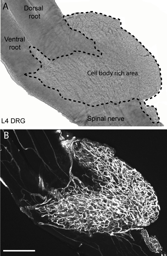
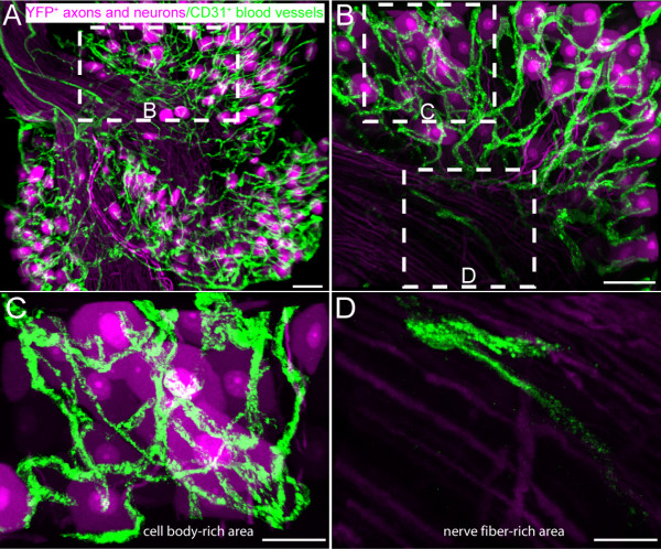
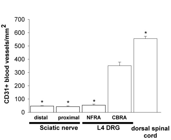
Although a variety of industrial chemicals, as well as several chemotherapeutic agents used to treat cancer or HIV, preferentially induce a peripheral sensory neuropathy what remains unclear is why these agents induce a sensory vs. a motor or mixed neuropathy. Previous studies have shown that the endothelial cells that vascularize the dorsal root ganglion (DRG), which houses the primary afferent sensory neurons, are unique in that they have large fenestrations and are permeable to a variety of low and high molecular weight agents. In the present report we used whole-mount preparations, immunohistochemistry, and confocal laser scanning microscopy to show that the cell body-rich area of the L4 mouse DRG has a 7 fold higher density of CD31+ capillaries than cell fiber rich area of the DRG or the distal or proximal aspect of the sciatic nerve. This dense vascularization, coupled with the high permeability of these capillaries, may synergistically contribute, and in part explain, why many potentially neurotoxic agents preferentially accumulate and injure cells within the DRG. Currently, cancer survivors and HIV patients constitute the largest and most rapidly expanding groups that have chemically induced peripheral sensory neuropathy. Understanding the unique aspects of the vascularization of the DRG and closing the endothelial fenestrations of the rich vascular bed of capillaries that vascularize the DRG before intravenous administration of anti-neoplastic or anti-HIV therapies, may offer a mechanism based approach to attenuate these chemically induced peripheral neuropathies in these patients.
Figures
References
-
- Le Quesne PM. Neuropathy due to drugs. In: Dyck PF, Thomas PK, Lambert EH, editor. Peripheral Neuropathy. Philadelphia , Saunders; 1975.
-
- Cho ES, Schaumburg HH, Spencer PS. Adriamycin produces ganglioradiculopathy in rats. J Neuropathol Exp Neurol. 1977;36:907–915. - PubMed
-
- Hopkins A. Toxic neuropathy due to industrial agents. In: Dyck PF, Thomas PK, Lambert EH, editor. Peripheral Neuropathy. Philadelphia , Saunders; 1975.
-
- Grant MW. Toxicology of the eye. In: Thomas CC, editor. Toxicology of the eye. Illinois , Springfield; 1974. p. 96.
Publication types
MeSH terms
Substances
Grants and funding
LinkOut - more resources
Full Text Sources
Medical
