. 2008 May;179(5):1850-6.
doi: 10.1016/j.juro.2008.01.047. Epub 2008 Mar 18.

Urine markers do not predict biopsy findings or presence of bladder ulcers in interstitial cystitis/painful bladder syndrome

Affiliations

Urine markers do not predict biopsy findings or presence of bladder ulcers in interstitial cystitis/painful bladder syndrome

Deborah R Erickson et al. J Urol. 2008 May.

Abstract

Purpose: We tested for associations between urine markers, bladder biopsy features and bladder ulcers in interstitial cystitis/painful bladder syndrome.

Materials and methods: Subjects were 72 patients with interstitial cystitis/painful bladder syndrome undergoing bladder distention and biopsy. Urine was collected before the procedure. Urine marker levels were correlated with biopsy and cystoscopic findings. Patients with no previous interstitial cystitis/painful bladder syndrome treatments (47) were analyzed separately from previously treated patients (25).

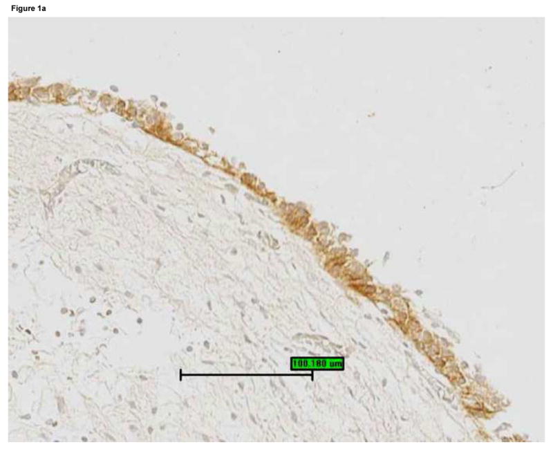
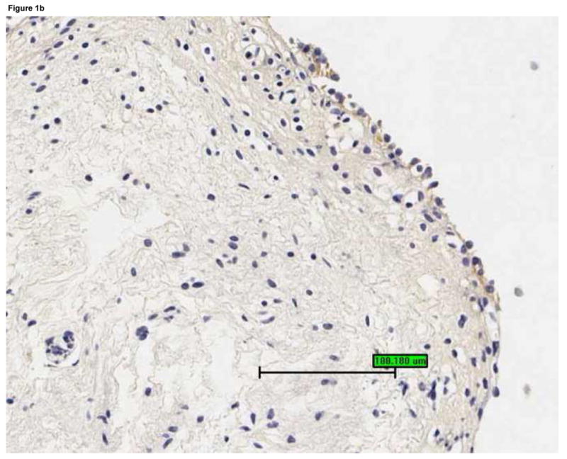
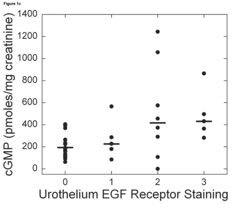
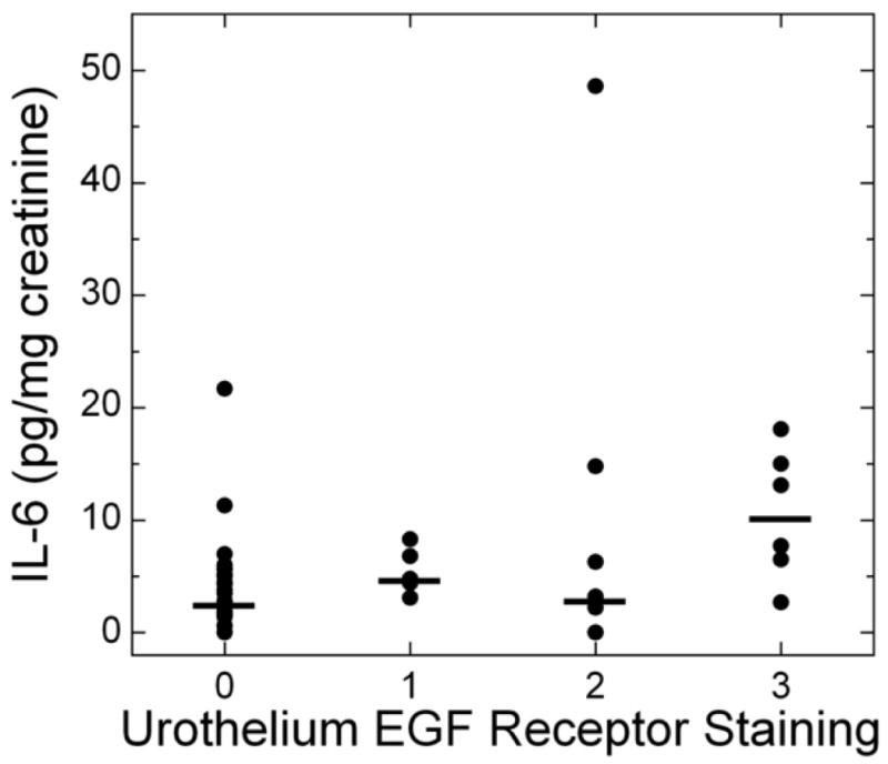
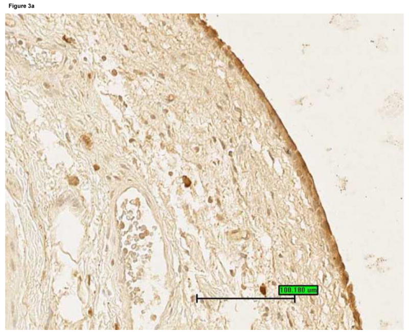
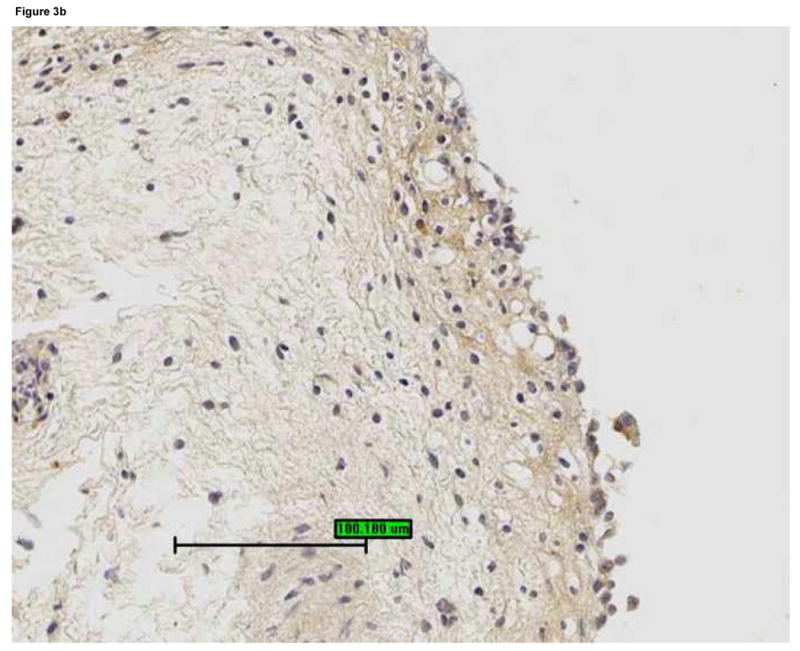
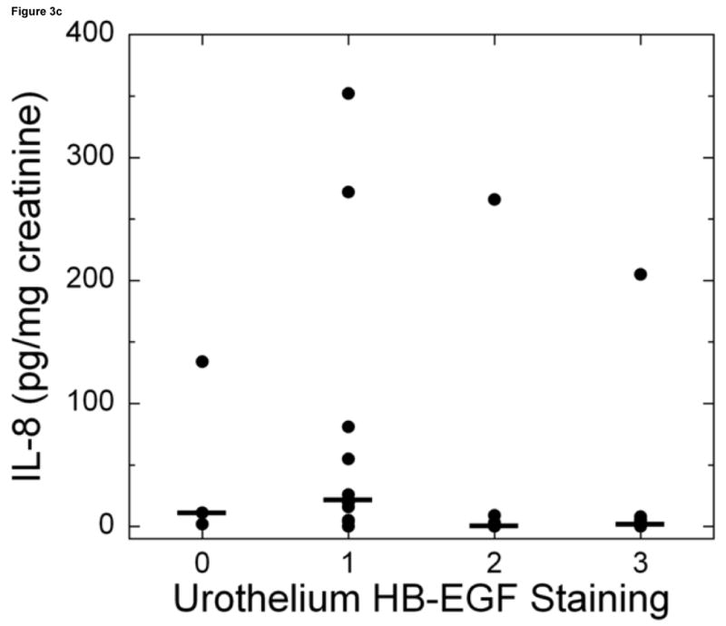
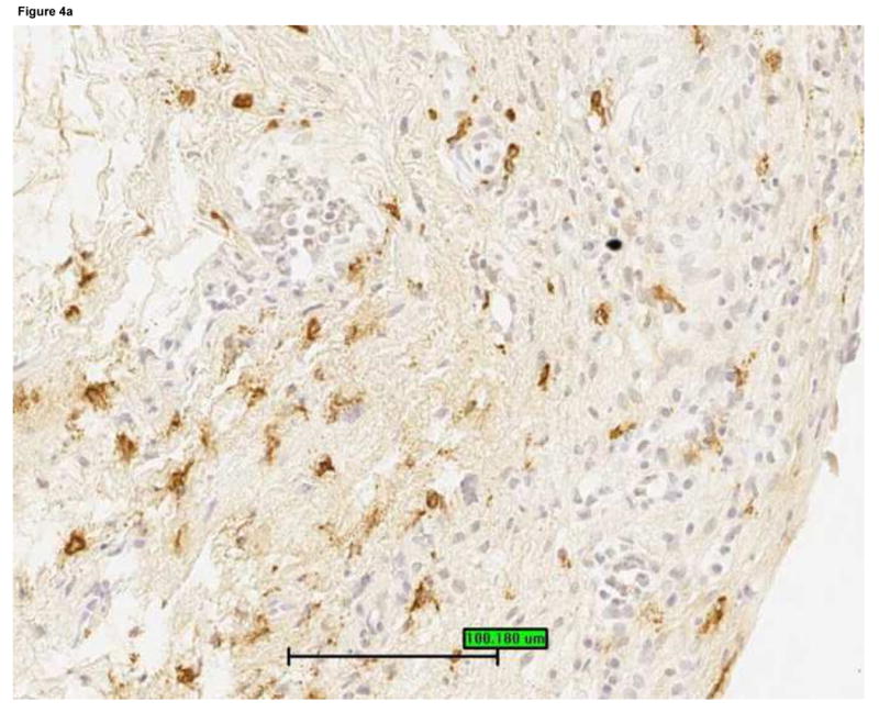
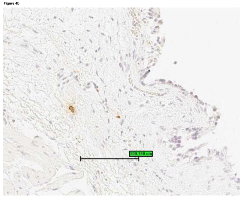
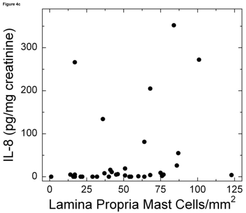
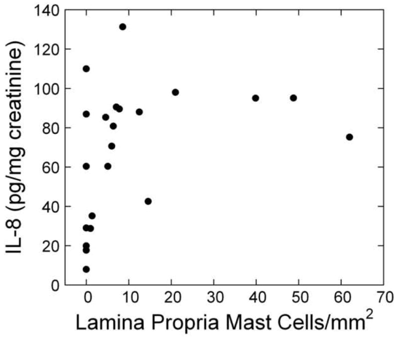
Results: For untreated patients urine interleukin-6 and cyclic guanosine monophosphate were associated with urothelial epidermal growth factor receptor staining (for interleukin-6 r = 0.29; 95% CI 0.07, 0.51; p = 0.01 and for cyclic guanosine monophosphate r = 0.34; 95% CI 0.13, 0.55; p = 0.002). Urine interleukin-8 was negatively associated with urothelial heparin-binding epidermal growth factor-like growth factor staining (r = -0.34; 95% CI -0.55, -0.12; p = 0.002) and positively associated with lamina propria mast cell count (r = 0.29; 95% CI 0.06, 0.52; p = 0.01). The latter association also was seen in treated patients (r = 0.46; 95% CI 0.20, 0.73; p <0.001). None of the urine markers was significantly different for ulcer vs nonulcer groups. All of the patients with ulcer had extensive inflammation on bladder biopsy including severe mononuclear cell infiltration, moderate or strong interleukin-6 staining in the urothelium and lamina propria, and leukocyte common antigen staining in more than 10% of the lamina propria. However, these features also were seen in 24% to 76% of the patients without ulcer.

Conclusions: Overall urine markers did not associate robustly with biopsy findings. The strongest association was a positive association between urine interleukin-8 levels and bladder mast cell count. Patients with ulcer consistently had bladder inflammation but the cystoscopic finding of ulcers was not a sensitive indicator of inflammation on bladder biopsy.


Figures

Figure 1
Figure 1
Bladder biopsies were stained with anti-EGFR primary antibody, followed by a standard streptavidin method. EGFR staining was graded semiquantitatively from 0 (none) to 3 (intense). On each biopsy the urothelium and lamina propria were graded separately. a: Grade 3 staining for EGFR in the urothelium (magnification 200×). b: Grade 1 staining for EGFR in the urothelium (magnification 200×). c: Urothelial EGFR staining intensity was significantly associated with urine cGMP levels in untreated IC/PBS patients. Urine cGMP was analyzed by radioimmunoassay and normalized to urine creatinine. Bars represent median values for cGMP in each staining category.
Figure 1
Figure 1
Bladder biopsies were stained with anti-EGFR primary antibody, followed by a standard streptavidin method. EGFR staining was graded semiquantitatively from 0 (none) to 3 (intense). On each biopsy the urothelium and lamina propria were graded separately. a: Grade 3 staining for EGFR in the urothelium (magnification 200×). b: Grade 1 staining for EGFR in the urothelium (magnification 200×). c: Urothelial EGFR staining intensity was significantly associated with urine cGMP levels in untreated IC/PBS patients. Urine cGMP was analyzed by radioimmunoassay and normalized to urine creatinine. Bars represent median values for cGMP in each staining category.
Figure 2
Figure 2
Urine IL-6 levels vs. EGF receptor staining in the urothelium for untreated patients. Bladder biopsies were stained with anti-EGFR primary antibody followed by a standard streptavidin method. EGFR staining was graded semiquantitatively from 0 (none) to 3 (intense). (For examples of strong and weak EGFR staining, see Figure 1). On each biopsy the urothelium and lamina propria were graded separately. Urothelial EGFR staining intensity was significantly associated with urine IL-6 levels in untreated IC/PBS patients. Urine IL-6 was analyzed by ELISA and normalized to urine creatinine. Bars represent median values for IL-6 in each staining category.
Figure 3
Figure 3
Bladder biopsies were stained with anti-HB-EGF primary antibody followed by a standard streptavidin method. HB-EGF staining was graded semiquantitatively from 0 (none) to 3 (intense). On each biopsy the urothelium and lamina propria were graded separately. a: Grade 3 staining for HB-EGF in the urothelium and lamina propria (magnification 200×). b: Grade 1 staining for HB-EGF in the urothelium and lamina propria (magnification 200×). c: Urothelial HB-EGF staining intensity was significantly associated with urine IL-8 levels in untreated IC/PBS patients. Urine IL-8 was analyzed by ELISA and normalized to urine creatinine. Bars represent median values for IL-8 in each staining category.
Figure 3
Figure 3
Bladder biopsies were stained with anti-HB-EGF primary antibody followed by a standard streptavidin method. HB-EGF staining was graded semiquantitatively from 0 (none) to 3 (intense). On each biopsy the urothelium and lamina propria were graded separately. a: Grade 3 staining for HB-EGF in the urothelium and lamina propria (magnification 200×). b: Grade 1 staining for HB-EGF in the urothelium and lamina propria (magnification 200×). c: Urothelial HB-EGF staining intensity was significantly associated with urine IL-8 levels in untreated IC/PBS patients. Urine IL-8 was analyzed by ELISA and normalized to urine creatinine. Bars represent median values for IL-8 in each staining category.
Figure 4
Figure 4
Mast cells were detected by staining bladder biopsies with anti-tryptase primary antibody followed by a standard streptavidin method. An optical reticle was used to count mast cells in the urothelium, lamina propria and detrusor. a: Brisk mastocytosis in the lamina propria (magnification 200×). b: Minimal mastocytosis in the lamina propria (magnification 200×). c: Scatter plot of urine IL-8 levels vs. mast cell count in the lamina propria for untreated IC/PBS patients. Lamina propria mast cell count was significantly associated with urine IL-8, which was analyzed by ELISA and normalized to urine creatinine.
Figure 4
Figure 4
Mast cells were detected by staining bladder biopsies with anti-tryptase primary antibody followed by a standard streptavidin method. An optical reticle was used to count mast cells in the urothelium, lamina propria and detrusor. a: Brisk mastocytosis in the lamina propria (magnification 200×). b: Minimal mastocytosis in the lamina propria (magnification 200×). c: Scatter plot of urine IL-8 levels vs. mast cell count in the lamina propria for untreated IC/PBS patients. Lamina propria mast cell count was significantly associated with urine IL-8, which was analyzed by ELISA and normalized to urine creatinine.
Figure 5
Figure 5
Scatter plot of urine IL-8 levels vs. mast cell count in the lamina propria for chronically treated IC/PBS patients. Mast cells were detected by staining bladder biopsies with anti-tryptase primary antibody followed by a standard streptavidin method. An optical reticle was used to count mast cells in the urothelium, lamina propria and detrusor. (For examples of high and low mast cell counts, see Figure 4.) Lamina propria mast cell count was significantly associated with urine IL-8, which was analyzed by ELISA and normalized to urine creatinine.

Comment in

References

    1. Erickson DR, Tomaszewski JE, Kunselman AR, Bentley CM, Peters KM, Rovner ES, Demers LM, Wheeler MA, Keay SK. Do the National Institute of Diabetes and Digestive and Kidney Diseases cystoscopic criteria associate with other clinical and objective features of interstitial cystitis? J Urol. 2005;173:93. - PubMed
    1. Hanno PM, Landis JR, Matthews-Cook Y, Kusek J, Nyberg L the Interstitial Cystitis Database Study Group. The diagnosis of interstitial cystitis revisited: lessons learned from the National Institutes of Health Interstitial Cystitis Database Study. J Urol. 1999;161:553. - PubMed
    1. Erickson DR, Xie SX, Bhavanandan VP, Wheeler MA, Hurst RE, Demers LM, Kushner L, Keay SK. A comparison of multiple urine markers for interstitial cystitis. J Urol. 2002;167:2461. - PubMed
    1. Tomaszewski JE, Landis JR, Russack V, Williams TM, Wang LP, Hardy C, Brensinger C, Matthews YL, Abele ST, Kusek JW, Nyberg LM The Interstitial Cystitis Database Study Group. Biopsy features are associated with primary symptoms in interstitial cystitis: results from the interstitial cystitis database study. Urology. 2001;57(6 Suppl 1):67. - PubMed
    1. Peeker R, Fall M. Toward a precise definition of interstitial cystitis: further evidence of differences in classic and nonulcer disease. J Urol. 2002;167:2470. - PubMed

Publication types

MeSH terms