. 2008 Jun 5;375(2):464-79.
doi: 10.1016/j.virol.2008.02.020. Epub 2008 Mar 18.

Dephosphorylation of JC virus agnoprotein by protein phosphatase 2A: inhibition by small t antigen

Affiliations

Dephosphorylation of JC virus agnoprotein by protein phosphatase 2A: inhibition by small t antigen

Ilker K Sariyer et al. Virology. .

Abstract

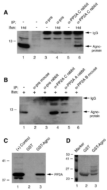
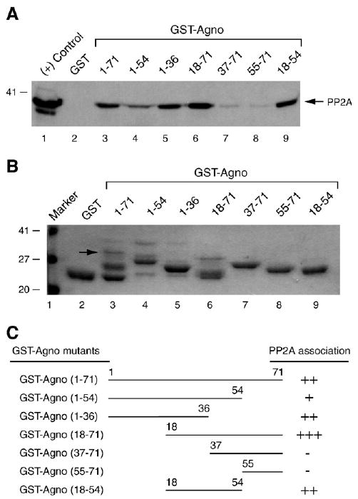
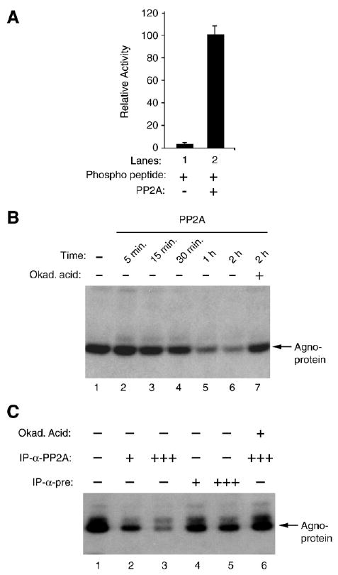
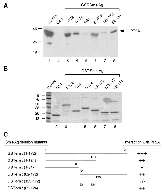
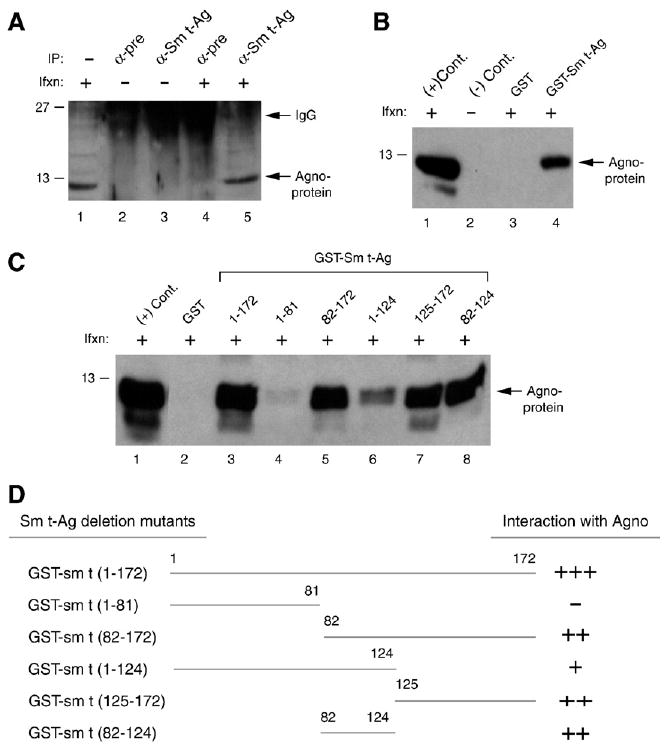
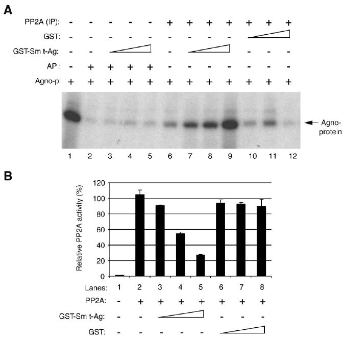
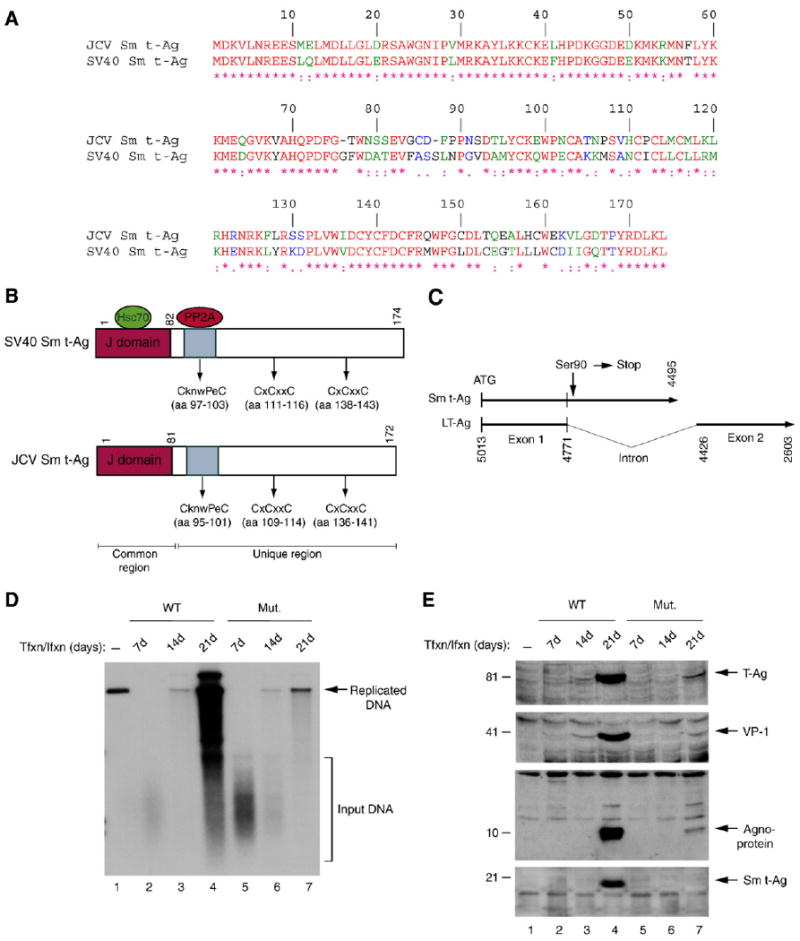
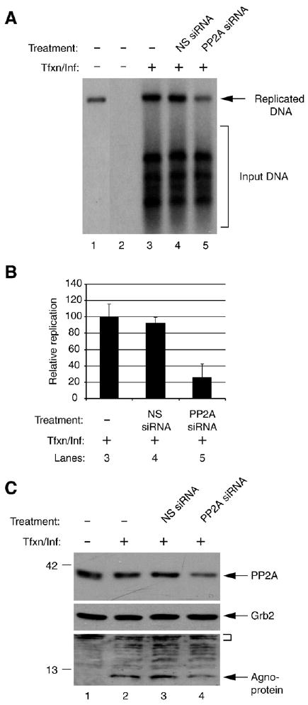
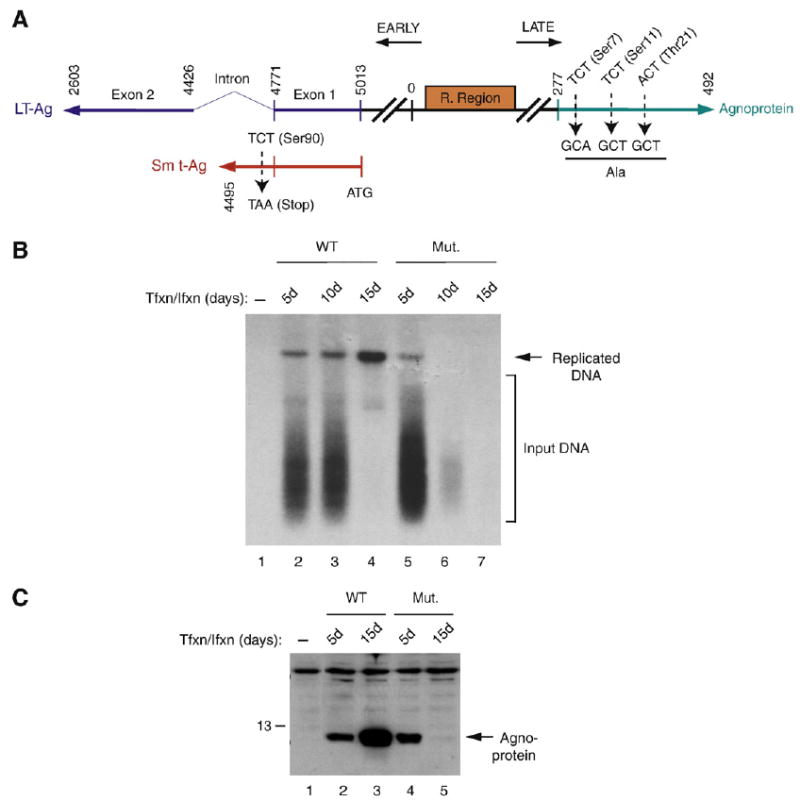
Previous studies have demonstrated that the JC virus (JCV) late regulatory protein agnoprotein is phosphorylated by the serine/threonine-specific protein kinase-C (PKC) and mutants of this protein at the PKC phosphorylation sites exhibit defects in the viral replication cycle. We have now investigated whether agnoprotein phosphorylation is regulated by PP2A, a serine/threonine-specific protein phosphatase and whether JCV small t antigen (Sm t-Ag) is involved in this regulation. Protein-protein interaction studies demonstrated that PP2A associates with agnoprotein and dephosphorylates it at PKC-specific sites. Sm t-Ag was also found to interact with PP2A and this interaction inhibited the dephosphorylation of agnoprotein by PP2A. The interaction domains of Sm t-Ag and agnoprotein with PP2A were mapped, as were the interaction domains of Sm t-Ag with agnoprotein. The middle portion of Sm t-Ag (aa 82-124) was found to be critical for the interaction with both agnoprotein and PP2A and the N-terminal region of agnoprotein for interaction with Sm t-Ag. To further understand the role of Sm t-Ag in JCV regulation, a stop codon was introduced at Ser90 immediately after splice donor site of the JCV early gene and the functional consequences of this mutation were investigated. The ability of this mutant virus to replicate was substantially reduced compared to WT. Next, the functional significance of PP2A in JCV replication was examined by siRNA targeting. Downregulation of PP2A caused a significant reduction in the level of JCV replication. Moreover, the impact of Sm t-Ag on agnoprotein phosphorylation was investigated by creating a double mutant of JCV, where Sm t-Ag stop codon mutant was combined with an agnoprotein triple phosphorylation mutant (Ser7, Ser11 and Thr21 to Ala). Results showed that double mutant behaves much like the triple phosphorylation mutant of agnoprotein during viral replication cycle, which suggests that agnoprotein might be an important target of Sm t-Ag with respect to the regulation of its phosphorylation. Collectively, these results suggest that there is an interplay between agnoprotein, Sm t-Ag and PP2A with respect to the regulation of JCV life cycle and this could be important for the progression of the JCV-induced disease, PML.


Figures

Fig. 1
Fig. 1
Agnoprotein interacts with PP2A. (A) Coimmunoprecipitation of agnoprotein with PP2A C subunit. Whole cell extracts (500 μg) were prepared from SVG-A cells [uninfected (−) or infected with JCV Mad-1] at the indicated days (d) post infection, subjected to immunoprecipitation (IP) using α-pre or α-PP2A C antibody (2 μg) as indicated. Immunocomplexes were then analyzed by Western blotting using α-agnoprotein antibody as described in Materials and methods. In lane 1, whole cell extract from SVG-A cells (20 μg) was loaded as a positive control for agnoprotein. IgG-labeled arrow points the light chain of the antibodies that were used for IP and detected by the secondary antibody. Ifxn denotes infection. (B) In parallel to the immunoprecipitation studies described in panel B, protein complexes were also precipitated (IP) with antibodies that are directed against A (α-PP2A A rabbit polyclonal, B (α-PP2A B mouse monoclonal) and C (α-PP2A C rabbit polyclonal) subunits of PP2A or either with normal mouse serum (α-pre M) or normal rabbit serum (α-pre R) as indicated using whole cell extracts from SVG-A cells prepared at 14 days post infection. Immunocomplexes were then analyzed by Western blotting using an anti-agno rabbit antibody. (C) GST pulldown assay. Whole cell extracts prepared from SVG-A cells were incubated either with GST or GST–Agnoprotein (Agno) (2 μg each), immobilized on GST–Sepharose beads. Columns were extensively washed with binding buffer and retained proteins were analyzed by Western blotting using α-PP2A C antibody as described in Materials and methods. In lane 1, whole cell extract from SVG-A cells (20 μg) was loaded as a positive control. (D) SDS-PAGE analysis of GST and GST–agnoprotein followed by coomassie staining.
Fig. 2
Fig. 2
Eighteen to thirty-six amino acid region of agnoprotein is critical for interaction with PP2A. (A) Bacterially produced GST or GST–agno fusion protein or agnoprotein deletion mutants that were fused to GST were immobilized on GST–Sepharose beads (2 μg each) and incubated with whole cell extracts (300 μg) prepared from SVG-A cells overnight at 4 ° by racking. Unbound proteins were washed and bound proteins were analyzed by Western blotting using α-PP2A C antibody. In lane 1, whole cell extract was directly loaded on the gel as a positive control (20 μg). (B) SDS-PAGE analysis of GST, GST–Agno full-length and GST–Agno deletion mutants. Position of the GST–Agno full length was indicated by an arrow. (C) Summary of the results obtained from in vitro mapping assays. The relative binding activity of agnoprotein and its deletion mutants is represented by + or − signs. +++, very strong binding; ++, strong binding; +, binding; and −, no binding.
Fig. 3
Fig. 3
PP2A dephosphorylates agnoprotein. (A) Enzymatic activity of purified PP2A on authentic substrate was measured according to manufacturer's recommendations (Upstate) and expressed as relative activity compared to without enzyme in the reaction. (B) Time course activity of PP2A on phosphorylated agnoprotein. PKC-phosphorylated GST–agnoprotein was equally distributed into reaction tubes, subjected to dephosphorylation by purified PP2A at different time points and analyzed by autoradiography. Okadaic acid was used as a specific inhibitor of PP2A in the reaction (lane 7). (C) PKC-phosphorylated GST–agnoprotein was also subjected to dephosphorylation by immunoprecipitated-PP2A and analyzed by autoradiography as described in Materials and methods. PP2A was immunoprecipitated (IP) from whole cell extracts prepared from SVG-A cells (500 μg) using either α-pre (2 μg) or α-PP2A C antibody (2 μg) prior to dephosphorylation reaction. Immunoprecipitated PP2A was split into 4 equal portions. One (+) or three (+++) portions of immunoprecipitated PP2A were used in the reactions as indicated. In lane 6, okadaic acid (Okad. Acid) was used to inhibit the dephosphorylation reaction.
Fig. 4
Fig. 4
The PP2A interaction domain of the Sm t-Ag localizes to its middle portion. (A) Sm t-Ag and its deletion mutants were expressed in bacteria as fusion proteins and immobilized on glutathione-S-transferase (GST) beads. Whole cell lysates prepared from U-87MG cells were incubated with either GST–Sm t-Ag or its deletion mutants for 2 h. The columns were extensively washed with binding buffer as described in Materials and methods and the proteins retained in the column were fractionated by SDS-10% PAGE and analyzed by Western blotting with an antibody directed against PP2A. (B) Analysis of GST, GST–Sm t-Ag and the deletion mutants of Sm t-Ag by SDS-12% PAGE. (C) Schematic representation of Sm t-Ag and its deletion mutants. The relative binding activity of Sm t-Ag and its deletion mutants is represented by + or − signs. +++, very strong binding; ++, strong binding; +, binding; +/−, weak binding and −, no binding.
Fig. 5
Fig. 5
The C-terminal portion of Sm t-Ag is important for agnoprotein interaction. (A) Agnoprotein coimmunoprecipitates with Sm t-Ag. Whole cell extracts (500 μg) prepared from SVG-A cells infected (Ifxn) or uninfected with JCV Mad-1 were subjected to immunoprecipitation (IP) using α-pre rabbit and α-Sm t-Ag antibody and analyzed by Western blotting using α-agno antibody. In lane 1, whole cell extracts from infected cells were loaded as a positive control. (B) GST pulldown assay. Whole cell extracts, prepared from SVG-A cells, infected with JCV Mad-1 were incubated with either GST or GST–Sm t-Ag, immobilized on Glutathione–Sepharose beads. Bound proteins were analyzed by Western blotting using an α-agno antibody as described for Fig. 2. In lane 1, whole cell extract (20 μg) from infected cells was loaded as a positive (+) control. In lane 2, whole cell extract (20 μg) from uninfected cells was loaded as a negative (−) control. (C) The agnoprotein interaction domain of Sm t-Ag maps to the C-terminal portion of the protein. GST pulldown assay was carried out as described for panel B. (D) Summary of the results from in vitro mapping assays as described for Fig. 4C.
Fig. 6
Fig. 6
Sm t-Ag inhibits agnoprotein dephosphorylation by PP2A. (B) Full-length agnoprotein was expressed in bacteria as a GST fusion protein (GST–Agno) and purified its homogeneity and phosphorylated by PKC as described previously (Sariyer et al., 2006). PKC-phosphorylated agnoprotein was subjected to dephosphorylation by PP2A, fractionated on SDS-PAGE and analyzed by autoradiography as described in Materials and methods. In lane 2, alkaline phosphatase (AP) was used as a control to dephosphorylate agnoprotein (Sadowska et al., 2003). In lanes 3–5, an increasing amount of GST–Sm t-Ag (0.5, 1.0 and 1.5 μg/lane respectively) was added to the reaction. Similarly, in lanes 7–9, GST–Sm t-Ag was also added into the reaction in increasing amount as described for lanes 3–5. In lane 10–12, an increasing amount of GST (0.5, 1.0 and 1.5 μg/lane respectively) alone was used. In lane 1, phosphorylated GST–agnoprotein was loaded as a positive control. (B) JCV Sm t-Ag inhibits PP2A activity. Inhibition reaction is described in Materials and methods in detail. Briefly, GST and GST Sm t-Ag was expressed in bacteria and purified to their homogeneity. Both proteins were added into reaction in increasing amounts (0.5. 1.0 and 1.5 μg/lane) as indicated and the amount of phosphate released from substrate was colorimetrically determined at 600 nm wavelengths. The enzyme activity was expressed in “relative PP2A activity” in percent.
Fig. 7
Fig. 7
Viral replication and gene expression were substantially down-regulated in the absence of full-length Sm t-Ag expression. (A) Amino acid alignment of JCV Sm t-Ag and SV40 Sm t-Ag. (B) Schematic comparison of SV40 Sm t-Ag regions with that of JCV Sm t-Ag. The position of J domains, PP2A binding domains and cystein clusters are indicated. (C) Schematic representation of JCV Mad-1 early coding region, where Ser90 (TCT) of Sm t-Ag was converted into a stop codon as described in Materials and methods. Numbering is according to JCV Mad-1 strain (GenBank # NC_001699, formerly J02226). (D) Dpn I assay. Primary human fetal glial cells (PHFG) (4 million cells/75 cm2 flask) were transfected/infected either with WT (5 μg) or Mut genome (5 μg) by lipofectin method. At 7d, 14d and 21d posttransfection, low molecular weight DNA was isolated (Ziegler et al., 2004) and analyzed by Dpn I assay (Hirt, 1976). (E) In parallel, either nuclear or cytoplasmic extracts were prepared from transfectants and analyzed by Western blotting using anti-LT-Ag (PAb-2000), anti-VP-1 (PAB597, a gift from Dr. W. Atwood), anti-agnoprotein and anti-Sm t-Ag antibodies as indicated.
Fig. 8
Fig. 8
Effect of PP2A inhibition on JCV replication and gene expression. (A) Inhibition of PP2A negatively affects JCV replication. SVG-A cells. 2 × 10⁵ cells per 35-mm tissue culture dish were transfected/infected with JCV genome (2 μg/plate) in the presence and absence of 50 pmol of non-targeting siRNA or Smartpool PP2AC siRNA (PP2AC siRNA, Dharmacon, Lafayette, CO). At the 48 h post infection, cells were trypsinized and split into two equal portions. One half of the samples were used to prepare low molecular weight DNA for Dpn I assay and the other portion was used to prepare whole cell extracts for Western blotting. DNA was digested with both Bam HI and Dpn I and analyzed by Southern blotting using labeled JCV genomic DNA as probe. Dpn I enzyme digests only transfected (input) DNA and leaves intact the newly replicated DNA. In lane 1, plasmid containing the Mad-1 genome was digested with Bam HI only and 0.1 ng of it was run on the gel as a positive control. In lane 2, the DNA sample from untransfected/uninfected cells were processed as in lanes 3–5 and was loaded as a negative control. The arrow points to the replicated DNA and the bracket indicates the input DNA. Tfxn/Inf denotes transfection/infection, i.e., introduction of the Mad-1 JCV genome into cells by transfection. (B) Quantitative representation of the results from panel A. The bands corresponding to the replicated DNA was quantitated in each lane (lanes 3–5) by densitometry and normalized relative to the band intensity on lane 3. The standard deviations of the data are presented as error bars (C) Western blot analysis of the whole cell extracts prepared in parallel to the replication assay in panel A, using specific antibodies against agnoprotein, PP2AC and Grb2. In lane 1, sample from untransfected/uninfected cells was loaded as a negative control. The small bracket denotes the non-specific bands that indicate the equal protein loading for agnoprotein. Grb2 was probed as a loading control for PP2AC.
Fig. 9
Fig. 9
Agnoprotein triple phosphorylation mutant is resistant to a mutation in Sm t-Ag. (A) Schematic representation of a double mutant of JCV Mad-1 strain on Sm t-Ag and agnoprotein. On agnoprotein, Ser7, Ser11 and Thr21 were converted into Ala. On Sm t-Ag, Ser90 was converted into a stop codon. (B). Analysis of the growth properties of JCV WT Mad-1 and double mutant by Dpn I assay. SVG-A cells (5 million cells/75 cm2 flask) were transfected/infected either with WT (5 μg) or Mut genome (5 μg) by lipofectin method. At 5d, 10d and 15d posttransfection, low molecular weight DNA was isolated (Ziegler et al., 2004) and analyzed by Dpn I assay (Hirt, 1976). (C) In parallel, cytoplasmic extracts were prepared from transfectants and analyzed by Western blotting using anti-agnoprotein antibody.

Similar articles

Cited by

References

    1. Akan I, Sariyer IK, Biffi R, Palermo V, Woolridge S, White MK, Amini S, Khalili K, Safak M. Human polyomavirus JCV late leader peptide region contains important regulatory elements. Virology. 2006;349(1):66–78. - PubMed
    1. Ali SH, DeCaprio JA. Cellular transformation by SV40 large Tantigen: interaction with host proteins. Semin Cancer Biol. 2001;11(1):15–23. - PubMed
    1. Arroyo JD, Hahn WC. Involvement of PP2A in viral and cellular transformation. Oncogene. 2005;24(52):7746–7755. - PubMed
    1. Berger JR, Concha M. Progressive multifocal leukoencephalopathy: the evolution of a disease once considered rare. J Neurovirology. 1995;1(1):5–18. - PubMed
    1. Boutros R, Lobjois V, Ducommun B. CDC25 phosphatases in cancer cells: key players? Good targets? Nat Rev Cancer. 2007;7(7):495–507. - PubMed

Publication types