

Review
doi: 10.1016/j.tim.2008.02.001.
Epub 2008 Mar 18.
Staying alive: bacterial inhibition of apoptosis during infection
Affiliations
- PMID: 18353648
- PMCID: PMC2746948
- DOI: 10.1016/j.tim.2008.02.001
Item in Clipboard
Review
Staying alive: bacterial inhibition of apoptosis during infection
Trends Microbiol.
2008 Apr.
Abstract
The ability of bacterial pathogens to inhibit apoptosis in eukaryotic cells during infection is an emerging theme in the study of bacterial pathogenesis. Prevention of apoptosis provides a survival advantage because it enables the bacteria to replicate inside host cells. Bacterial pathogens have evolved several ways to prevent apoptosis by protecting the mitochondria and preventing cytochrome c release, by activating cell survival pathways, or by preventing caspase activation. This review summarizes the most recent work on bacterial anti-apoptotic strategies and suggests new research that is necessary to advance the field.
Figures
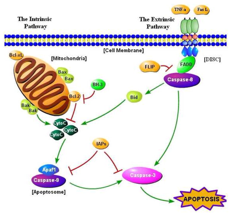
The apoptosis pathway. Apoptosis is activated by intrinsic and extrinsic pathways. In the intrinsic pathway, certain apoptotic stimuli alter the normal status of the Bcl-2 family of proteins (Box 1), which leads to permeabilization of the mitochondrial membrane. Under normal circumstances, the pro-survival proteins of the Bcl-2 family protect the mitochondrial membrane. Once cytochrome c (CytoC) is released into the cytosol, it binds to the apoptosome [1], a complex of proteins made up of the apoptosis activating factor-1 (Apaf1) protein and the initiator caspase-9. A morphological change in the apoptosome results from cytochrome c binding, which causes caspase-9 to become activated. Caspase-9 activates the effector caspase-3, leading to apoptosis [1]. Caspase-3 is known as the ‘executioner caspase’, because it activates or cleaves various protein targets, which is detrimental to the cell and results in death [1]. Activation of caspase-9 and caspase-3 are normally inhibited by a family of proteins known as the inhibitor of apoptosis proteins (IAPs), and XIAP (X-linked IAP) is the most potent IAP [1]. In the extrinsic pathway, ligands such as Fas ligand (FasL) or tumor necrosis factor α (TNF-α) bind to death receptors on the membrane of the host cell. Trimerization of the death receptors follows, forming what is known as the death-inducing signaling complex (DISC), which includes the Fas-associated death domain (FADD) adaptor protein, the Flice-like inhibitory protein (FLIP), and procaspase-8 [2]. Caspase-8 is activated, which in turn directly activates caspase-3 [1]. In addition, caspase-8 activates the pro-apoptotic protein Bid, which stimulates the intrinsic pathway to enhance the apoptotic signal, because Bid activation eventually leads to cytochrome c release [1].
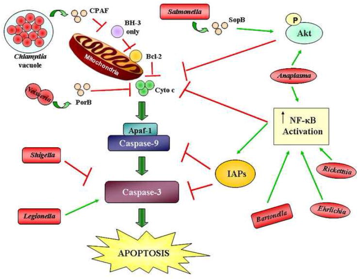
Mechanisms by which bacterial pathogens inhibit apoptosis at different points along the apoptotic pathway. Chlamydia secretes the chlamydial proteasome-like activity factor (CPAF) to inhibit and degrade the pro-apoptotic proteins with one BH3 domain. These pro-apoptotic proteins inhibit the pro-survival Bcl-2 proteins upon activation. The outer membrane protein porin PorB of Neisseria meningitidis prevents cytochrome c (CytoC) release. Salmonella secretes the effector SopB through a type III secretion system, resulting in the activation of the phosphatidylinositol 3-kinase/Akt (PI3K/Akt) pathway. This pathway prevents cytochrome c release. Anaplasma also activates the PI3K/Akt pathway in addition to activating nuclear factor kappa B (NF-κB). NF-κB prevents the release of cytochrome c and activates the inhibitor of apoptosis proteins (IAPs). Bartonella, Ehrlichia, and Rickettsia activate NF-κB as well. Shigella inhibits caspase-3 activation. Legionella directly activates caspase-3 to enhance infection, but inhibits apoptosis through NF-κB. Red lines indicate inhibition in the pathway while green arrows indicate activation. The bacterial proteins that specifically participate in apoptosis inhibition are shown, where known. The shapes of the bacteria, all shown in red, represent their morphology. P, phosphate.
Similar articles
-
Modification of host cell apoptosis by viral and bacterial pathogens.Int J Med Microbiol. 2001 Aug;291(3):197-207. doi: 10.1078/1438-4221-00125. Int J Med Microbiol. 2001. PMID: 11554560 Review.
-
Glutamate-induced apoptosis in primary cortical neurons is inhibited by equine estrogens via down-regulation of caspase-3 and prevention of mitochondrial cytochrome c release.BMC Neurosci. 2005 Feb 24;6:13. doi: 10.1186/1471-2202-6-13. BMC Neurosci. 2005. PMID: 15730564 Free PMC article.
-
Targeting of host organelles by pathogenic bacteria: a sophisticated subversion strategy.Nat Rev Microbiol. 2016 Jan;14(1):5-19. doi: 10.1038/nrmicro.2015.1. Epub 2015 Nov 23. Nat Rev Microbiol. 2016. PMID: 26594043 Review.
-
Live cell fluorescence microscopy to study microbial pathogenesis.Cell Microbiol. 2009 Apr;11(4):540-50. doi: 10.1111/j.1462-5822.2009.01283.x. Epub 2009 Jan 5. Cell Microbiol. 2009. PMID: 19134122 Free PMC article. Review.
-
Apaf-1/cytochrome c apoptosome: an essential initiator of caspase activation or just a sideshow?Cell Death Differ. 2003 Jan;10(1):16-8. doi: 10.1038/sj.cdd.4401166. Cell Death Differ. 2003. PMID: 12655291 Review. No abstract available.
Cited by
-
Burkholderia pseudomallei Differentially Regulates Host Innate Immune Response Genes for Intracellular Survival in Lung Epithelial Cells.PLoS Negl Trop Dis. 2016 Jul 1;10(7):e0004730. doi: 10.1371/journal.pntd.0004730. eCollection 2016 Jul. PLoS Negl Trop Dis. 2016. PMID: 27367858 Free PMC article.
-
Brucella dissociation is essential for macrophage egress and bacterial dissemination.Front Cell Infect Microbiol. 2014 Mar 5;4:23. doi: 10.3389/fcimb.2014.00023. eCollection 2014. Front Cell Infect Microbiol. 2014. PMID: 24634889 Free PMC article.
-
The role of DNA microarrays in Toxoplasma gondii research, the causative agent of ocular toxoplasmosis.J Ocul Biol Dis Infor. 2009 Dec 12;2(4):214-222. doi: 10.1007/s12177-009-9040-8. J Ocul Biol Dis Infor. 2009. PMID: 20157353 Free PMC article.
-
Human genome-wide RNAi screen for host factors that modulate intracellular Salmonella growth.PLoS One. 2012;7(6):e38097. doi: 10.1371/journal.pone.0038097. Epub 2012 Jun 11. PLoS One. 2012. PMID: 22701604 Free PMC article.
-
Identification of the essential Brucella melitensis porin Omp2b as a suppressor of Bax-induced cell death in yeast in a genome-wide screening.PLoS One. 2010 Oct 11;5(10):e13274. doi: 10.1371/journal.pone.0013274. PLoS One. 2010. PMID: 20949000 Free PMC article.
References
-
- Reed JC. Warner-Lambert/Parke-Davis Award lecture: Mechanisms of apoptosis. Am J Pathol. 2000;157:1415–1429. - PubMed
-
- Hyer ML, et al. The FLIP-side of Fas signaling. Clin Cancer Res. 2006;12:5929–5931. - PubMed
-
- Kroemer G, et al. Mitochondrial membrane permeabilization in cell death. Physiol Rev. 2007;87:99–163. - PubMed
-
- Hay S, Kannourakis G. A time to kill: viral manipulation of the cell death program. J Gen Virol. 2002;83:1547–1564. - PubMed
Publication types
MeSH terms
Substances
Grants and funding
LinkOut - more resources
Full Text Sources
Other Literature Sources
