Comparative Study
. 2008 May;30(2):255-69.
doi: 10.1016/j.nbd.2008.01.008. Epub 2008 Feb 16.

Prenatal choline supplementation attenuates neuropathological response to status epilepticus in the adult rat hippocampus

Affiliations
Comparative Study

Prenatal choline supplementation attenuates neuropathological response to status epilepticus in the adult rat hippocampus

Sarah J E Wong-Goodrich et al. Neurobiol Dis. 2008 May.

Abstract

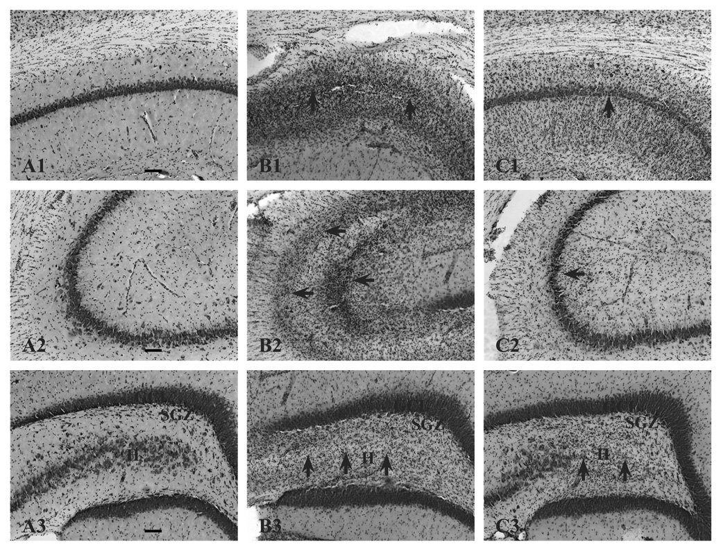
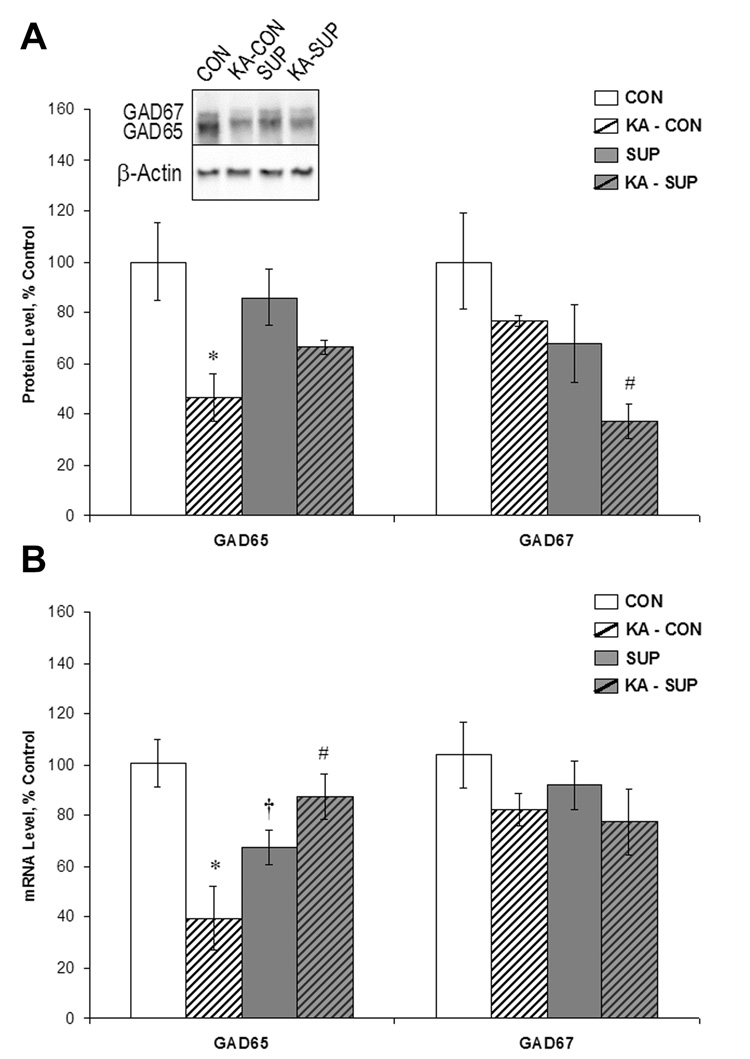
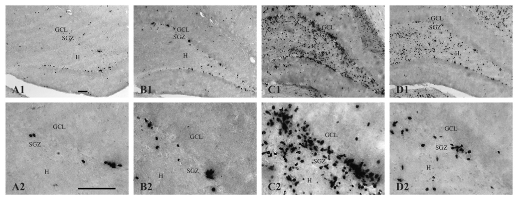
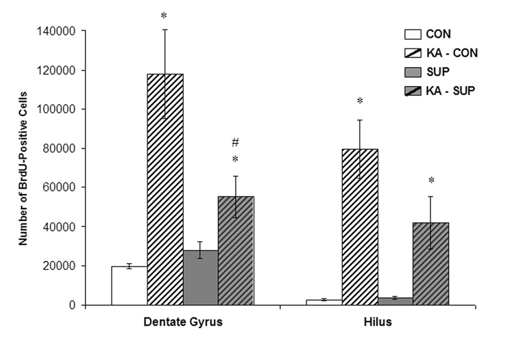
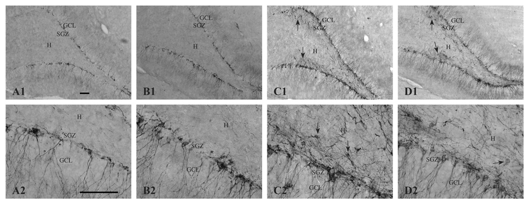
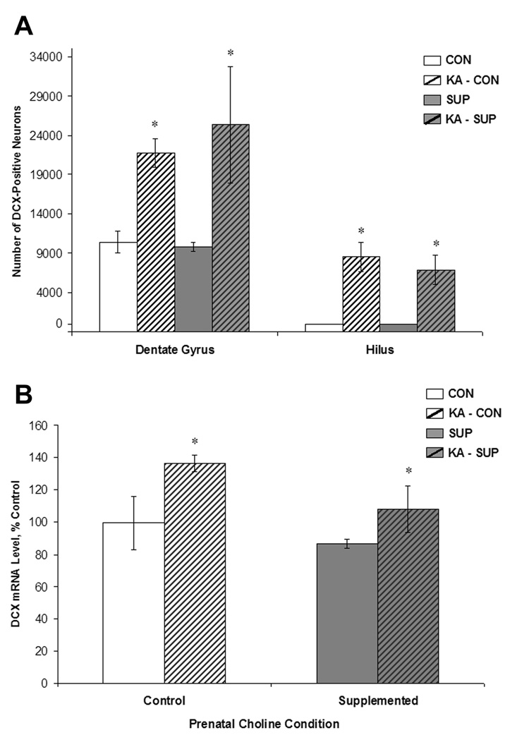
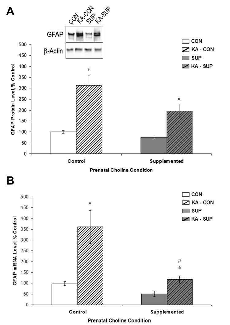
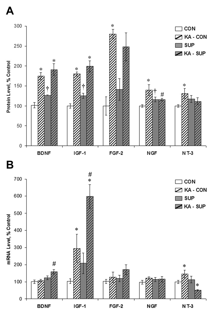
Prenatal choline supplementation (SUP) protects adult rats against spatial memory deficits observed after excitotoxin-induced status epilepticus (SE). To examine the mechanism underlying this neuroprotection, we determined the effects of SUP on a variety of hippocampal markers known to change in response to SE and thought to underlie ensuing cognitive deficits. Adult offspring from rat dams that received either a control or SUP diet on embryonic days 12-17 were administered saline or kainic acid (i.p.) to induce SE and were euthanized 16 days later. SUP markedly attenuated seizure-induced hippocampal neurodegeneration, dentate cell proliferation, and hippocampal GFAP mRNA expression levels, prevented the loss of hippocampal GAD65 protein and mRNA expression, and altered growth factor expression patterns. SUP also enhanced pre-seizure hippocampal levels of BDNF, NGF, and IGF-1, which may confer a neuroprotective hippocampal microenvironment that dampens the neuropathological response to and/or helps facilitate recovery from SE to protect cognitive function.


Figures

Figure 1
Figure 1
Histopathology of the hippocampus 16 days following KA-induced SE in CON (B) and SUP (C) rats. Panel A depicts representative sections of CA1 (A1), CA3 (A2), and dentate gyrus (A3) regions from an intact hippocampus from a saline-treated CON rat. Saline-treated SUP rats also did not show any lesions (histology data not shown). Note more severe degeneration (cell loss, disruption of cytoarchitecture, and gliosis) in KA-treated CON rats (B1, B2, B3). Areas of damage are indicated by arrows. Dentate hilar cell loss was observed in both CON (B3) and SUP rats (C3). Photomicrographs in each set were taken with a 10x objective. Bars indicate 50 µm. GCL, granule cell layer. SGZ, subgranular zone. H, hilus.
Figure 2
Figure 2
Comparison between CON (white bars) and SUP rats (grey bars) in GAD65 and GAD67 protein levels (A) and mRNA levels (B) (mean ± SEM percent of control levels) in the intact hippocampus (open bars) and 16 days following KA-induced SE (hatched bars). Protein levels were quantified using Western blot analysis (A). KA-treated CON rats showed a significant decrease in GAD65 protein and mRNA levels by more than half (p < .001), whereas GAD65 protein and mRNA levels did not significantly change in KA-treated SUP rats. GAD67 protein levels were reduced in both KA-treated CON and SUP rats, but GAD67 mRNA levels did not change in either group as a result of KA-induced SE. * statistically different from within-diet saline-treated group; # statistically different from KA-treated CON; † statistically different from saline-treated CON.
Figure 3
Figure 3
BrdU-immunopositive cells (i.e., newly generated cells) in the dentate gyrus and hilus 16 days following saline treatment (A, CON; B, SUP) or KA-induced SE (C, CON; D, SUP). Note that the number of BrdU-labeled cells significantly increased 16 days after SE for both diet groups, but that this seizure-induced proliferative response was markedly attenuated in SUP rats (D) in comparison to CON rats (C). Photomicrographs in the top row were taken with a 10x objective and the bottom row was taken with a 40x objective. Bars indicate 50 µm. GCL, granule cell layer. SGZ, subgranular zone. H, hilus.
Figure 4
Figure 4
Mean (±SEM) numbers of BrdU-labeled cells detected in the dentate gyrus and hilus of CON (white bars) and SUP (grey bars) rats 16 days following saline treatment (open bars) or KA-induced SE (hatched bars). SE significantly increased the number of BrdU-labeled cells in both CON and SUP rats (ps < .05), but this increase was significantly attenuated in SUP rats (ps < .05). * statistically different from within-diet saline-treated group; # statistically different from KA-treated CON.
Figure 5
Figure 5
DCX-immunopositive neurons (i.e., newly generated neurons) in the dentate gyrus and hilus 16 days following saline treatment (A, CON; B, SUP) or KA-induced SE (C, CON; D, SUP). The number of DCX-labeled cells significantly increased 16 days after SE for both CON (A, C) and SUP (B, D) rats. Note that in KA-treated rats of both diet groups (C, D), DCX-positive neurons were aberrantly located in the hilus and exhibited abnormal morphological features, such as horizontally oriented cell bodies and processes (indicated by arrows). Photomicrographs in the top row were taken with a 10x objective and the bottom row was taken with a 40x objective. Bars indicate 50 µm. GCL, granule cell layer. SGZ, subgranular zone. H, hilus.
Figure 6
Figure 6
Mean (±SEM) numbers of DCX-labeled cells (A) and mean (±SEM) mRNA expression levels (B) detected in the dentate gyrus and hilus of CON (white bars) and SUP (grey bars) rats 16 days following saline treatment (open bars) or KA-induced SE (hatched bars). KA-induced SE significantly increased the number of DCX-labeled neurons and levels of DCX mRNA in both CON and SUP rats (ps < .05). Prenatal choline supplementation did not alter the number of DCX-labeled neurons or levels of DCX mRNA in response to KA-induced SE. * statistically different from within-diet saline-treated group.
Figure 7
Figure 7
Comparison between CON (white bars) and SUP rats (grey bars) in GFAP protein levels (A) and mRNA levels (B) (mean ± SEM percent of control levels) in the intact hippocampus (open bars) and 16 days following KA-induced SE (hatched bars). GFAP protein levels were quantified using Western blot analysis (A). SE significantly increased GFAP protein and mRNA levels in both CON and SUP rats (ps < .05), but this increase was significantly attenuated in SUP rats. * statistically different from within-diet saline-treated group; # statistically different from KA-treated CON.
Figure 8
Figure 8
Comparison between CON (white bars) and SUP (grey bars) rats in growth factor protein (A) and mRNA (B) levels (mean ± SEM percent of control levels) in the intact hippocampus (open bars) and 16 days following KA-induced SE (hatched bars). Protein levels were quantified using ELISA. SE led to a significant increase in BDNF, IGF-1, FGF-2, NGF, and NT-3 protein levels in CON rats, but a significant increase in only BDNF and IGF-1 protein levels was noted for SUP rats. Baseline protein levels of BDNF, IGF-1, and NGF were significantly higher in SUP rats. SUP rats also exhibited significant increases in BDNF and IGF-1 mRNA and a decrease in NT-3 mRNA 16 d following SE, whereas CON rats showed SE-induced increases in IGF-1 and NT-3 mRNA. * statistically different from within-diet saline-treated group; # statistically different from KA-treated CON; † statistically different from saline-treated CON.

Similar articles

Cited by

References

    1. Anderson MF, Aberg MA, Nilsson M, Eriksson PS. Insulin-like growth factor-I and neurogenesis in the adult mammalian brain. Brain Res Dev Brain Res. 2002;134:115–122. - PubMed
    1. Ben-Ari Y, Represa A. Brief seizure episodes induce long-term potentiation and mossy fiber sprouting in the hippocampus. Trends Neurosci. 1990;13:312–318. - PubMed
    1. Bengzon J, Kokaia Z, Elmer E, Nanobashvili A, Kokaia M, Lindvall O. Apoptosis and proliferation of dentate gyrus neurons after single and intermittent limbic seizures. Proc Natl Acad Sci U S A. 1997;94:10432–10437. - PMC - PubMed
    1. Blusztajn JK. Choline, a vital amine. Science. 1998;281:794–795. - PubMed
    1. Blusztajn JK, Cermak JM, Holler T, Jackson DA. Imprinting of hippocampal metabolism of choline by its availability during gestation: implications for cholinergic neurotransmission. J Physiol Paris. 1998;92:199–203. - PubMed

Publication types