

Tensile properties of engineered cartilage formed from chondrocyte- and MSC-laden hydrogels
- PMID: 18353693
- PMCID: PMC2601559
- DOI: 10.1016/j.joca.2008.02.005
Tensile properties of engineered cartilage formed from chondrocyte- and MSC-laden hydrogels
Abstract
Objective: The objective of this study was to determine the capacity of chondrocyte- and mesenchymal stem cell (MSC)-laden hydrogel constructs to achieve native tissue tensile properties when cultured in a chemically defined medium supplemented with transforming growth factor-beta3 (TGF-beta3).
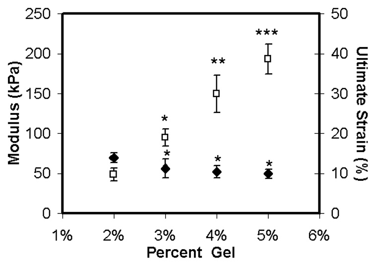
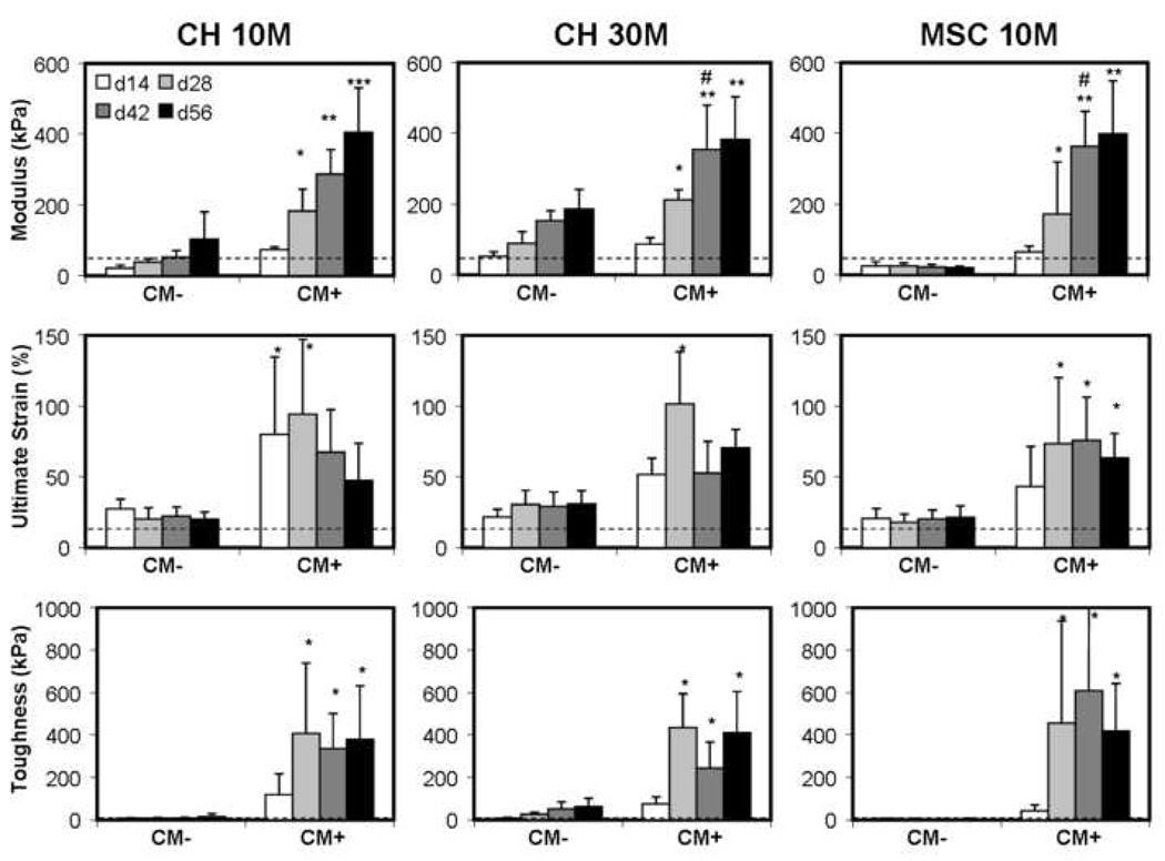
Design: Cell-laden agarose hydrogel constructs (seeded with bovine chondrocytes or MSCs) were formed as prismatic strips and cultured in a chemically defined serum-free medium in the presence or absence of TGF-beta3. The effects of seeding density (10 vs 30 million cells/mL) and cell type (chondrocyte vs MSC) were evaluated over a 56-day period. Biochemical content, collagenous matrix deposition and localization, and tensile properties (ramp modulus, ultimate strain, and toughness) were assessed biweekly.
Results: Results show that the tensile properties of cell-seeded agarose constructs increase with time in culture. However, tensile properties (modulus, ultimate strain, and toughness) achieved on day 56 were not dependent on either the initial seeding density or the cell type employed. When cultured in medium supplemented with TGF-beta3, tensile modulus increased and plateaued at a level of 300-400 kPa for each cell type and starting cell concentration. Ultimate strain and toughness also increased relative to starting values. Collagen deposition increased in constructs seeded with both cell types and at both seeding densities, with exposure to TGF-beta3 resulting in a clear shift toward type II collagen deposition as determined by immunohistochemical staining.
Conclusions: These findings demonstrate that the tensile properties, an important and often overlooked metric of cartilage development, increase with time in culture in engineered hydrogel-based cartilage constructs. Under the free-swelling conditions employed in the present study, tensile moduli and toughness did not match that of the native tissue, though significant time-dependent increases were observed with the inclusion of TGF-beta3. Of note, MSC-seeded constructs achieved tensile properties that were comparable to chondrocyte-seeded constructs, confirming the utility of this alternative cell source in cartilage tissue engineering. Further work, including both modulation of the chemical and mechanical culture environment, is required to optimize the deposition of collagen and its remodeling to achieve tensile properties in engineered constructs matching the native tissue.
Figures


Similar articles
-
Transient exposure to transforming growth factor beta 3 improves the mechanical properties of mesenchymal stem cell-laden cartilage constructs in a density-dependent manner.Tissue Eng Part A. 2009 Nov;15(11):3461-72. doi: 10.1089/ten.TEA.2009.0198. Tissue Eng Part A. 2009. PMID: 19432533 Free PMC article.
-
An in vitro and in vivo comparison of cartilage growth in chondrocyte-laden matrix metalloproteinase-sensitive poly(ethylene glycol) hydrogels with localized transforming growth factor β3.Acta Biomater. 2019 Jul 15;93:97-110. doi: 10.1016/j.actbio.2019.03.046. Epub 2019 Mar 23. Acta Biomater. 2019. PMID: 30914256 Free PMC article.
-
Functional cartilage repair capacity of de-differentiated, chondrocyte- and mesenchymal stem cell-laden hydrogels in vitro.Osteoarthritis Cartilage. 2014 Aug;22(8):1148-57. doi: 10.1016/j.joca.2014.05.019. Epub 2014 Jun 2. Osteoarthritis Cartilage. 2014. PMID: 24887551 Free PMC article.
-
A paradigm for functional tissue engineering of articular cartilage via applied physiologic deformational loading.Ann Biomed Eng. 2004 Jan;32(1):35-49. doi: 10.1023/b:abme.0000007789.99565.42. Ann Biomed Eng. 2004. PMID: 14964720 Review.
-
Mechanics and mechanobiology of mesenchymal stem cell-based engineered cartilage.J Biomech. 2010 Jan 5;43(1):128-36. doi: 10.1016/j.jbiomech.2009.09.018. Epub 2009 Oct 13. J Biomech. 2010. PMID: 19828149 Free PMC article. Review.
Cited by
-
Cartilage matrix formation by bovine mesenchymal stem cells in three-dimensional culture is age-dependent.Clin Orthop Relat Res. 2011 Oct;469(10):2744-53. doi: 10.1007/s11999-011-1869-z. Clin Orthop Relat Res. 2011. PMID: 21424832 Free PMC article.
-
Combined effects of oscillating hydrostatic pressure, perfusion and encapsulation in a novel bioreactor for enhancing extracellular matrix synthesis by bovine chondrocytes.Cell Tissue Res. 2017 Oct;370(1):179-193. doi: 10.1007/s00441-017-2651-7. Epub 2017 Jul 7. Cell Tissue Res. 2017. PMID: 28687928 Free PMC article.
-
Macromer density influences mesenchymal stem cell chondrogenesis and maturation in photocrosslinked hyaluronic acid hydrogels.Osteoarthritis Cartilage. 2009 Dec;17(12):1639-48. doi: 10.1016/j.joca.2009.07.003. Epub 2009 Jul 15. Osteoarthritis Cartilage. 2009. PMID: 19631307 Free PMC article.
-
Modular Orthopaedic Tissue Engineering With Implantable Microcarriers and Canine Adipose-Derived Mesenchymal Stromal Cells.Front Bioeng Biotechnol. 2020 Jul 22;8:816. doi: 10.3389/fbioe.2020.00816. eCollection 2020. Front Bioeng Biotechnol. 2020. PMID: 32775324 Free PMC article.
-
Tissue-engineered articular cartilage exhibits tension-compression nonlinearity reminiscent of the native cartilage.J Biomech. 2013 Jul 26;46(11):1784-91. doi: 10.1016/j.jbiomech.2013.05.017. Epub 2013 Jun 21. J Biomech. 2013. PMID: 23791084 Free PMC article.
References
-
- Guilak F, Sah RL, Setton LA. Physical regulation of cartilage metabolism. In: Mow VC, Hayes WC, editors. Basic orthopaedic biomechanics. Philadelphia: Lippincott-Raven; 1997. pp. 179–207.
-
- Mow VC, Ratcliffe A, Chern KY, Kelly MA. Structure and function relationships of the menisci of the knee. In: Mow VC, Arnoczky SP, Jackson DW, editors. Knee meniscus: basic and clinical foundations. New York: Raven Press, Ltd; 1992. pp. 37–57.
-
- Buckwalter JA, Martin J, Mankin HJ. Synovial joint degeneration and the syndrome of osteoarthritis. Instr Course Lect. 2000;49:481–489. - PubMed
-
- Hunziker EB. Articular cartilage repair: are the intrinsic biological constraints undermining this process insuperable? Osteoarthritis Cartilage. 1999;7:15–28. - PubMed
Publication types
MeSH terms
Substances
Grants and funding
LinkOut - more resources
Full Text Sources
