. 2008 May 9;283(19):13009-20.
doi: 10.1074/jbc.M801222200. Epub 2008 Mar 19.

AMP-activated protein kinase and p38 MAPK activate O-GlcNAcylation of neuronal proteins during glucose deprivation

Affiliations

AMP-activated protein kinase and p38 MAPK activate O-GlcNAcylation of neuronal proteins during glucose deprivation

Win D Cheung et al. J Biol Chem. .

Abstract

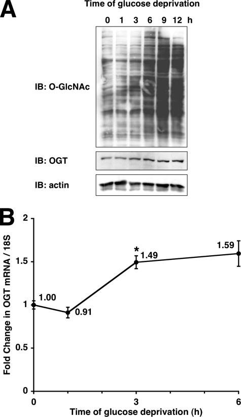
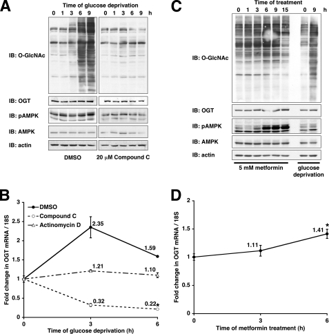
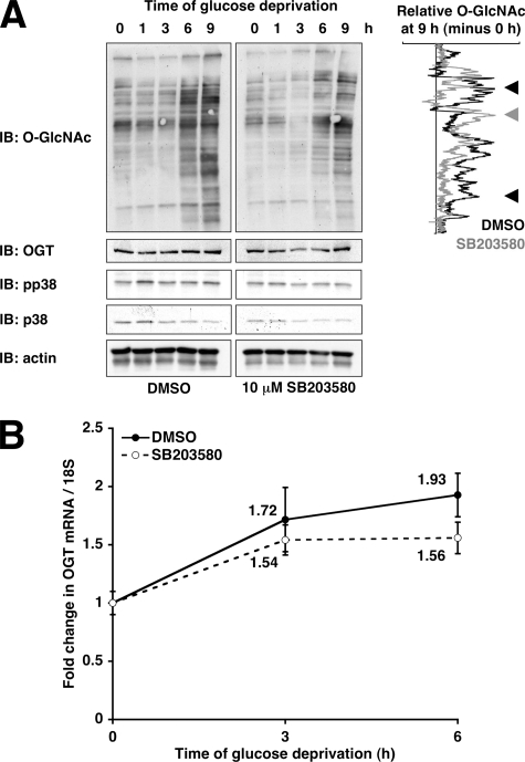
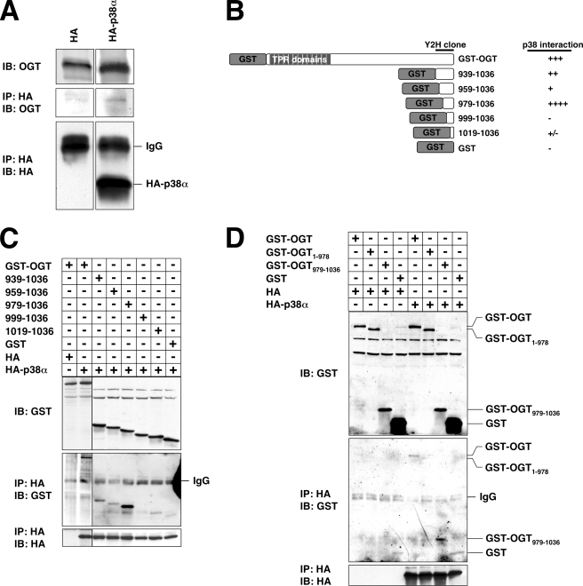
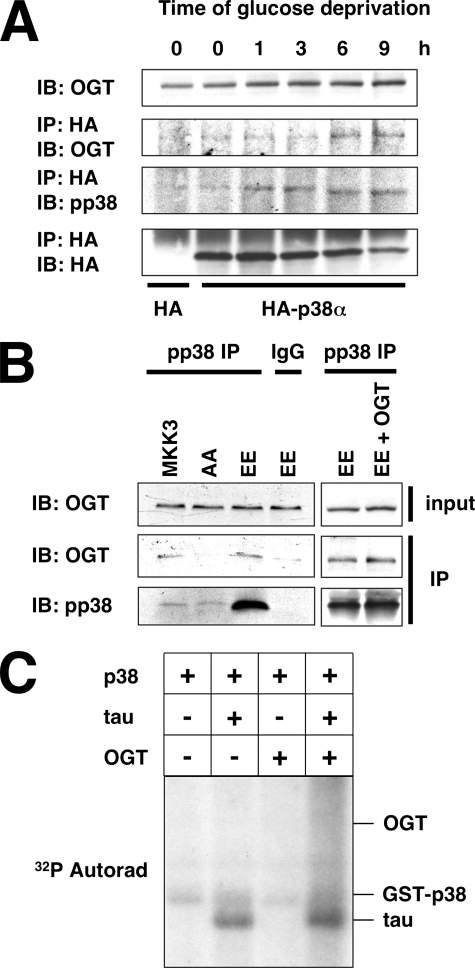
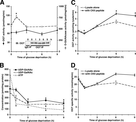
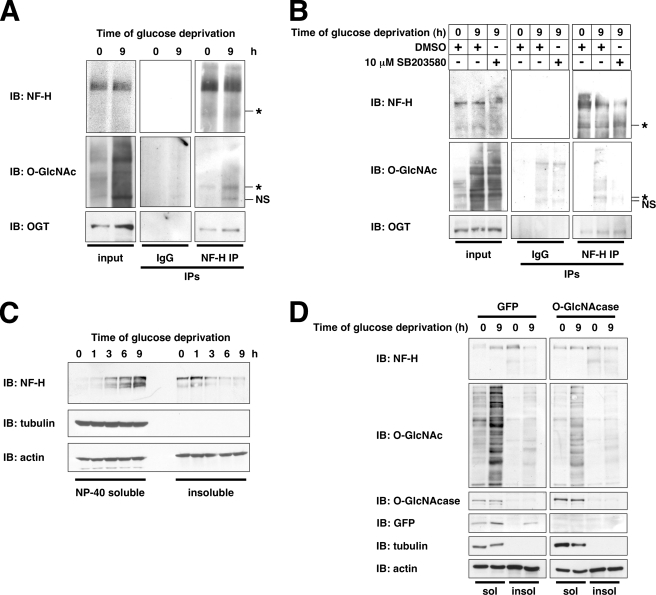
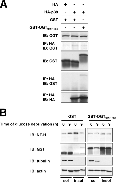
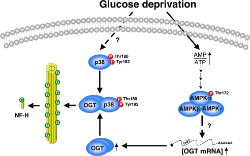
We have demonstrated previously that a wide array of stress signals induces O-GlcNAc transferase (OGT) expression and increases O-GlcNAcylation of many intracellular proteins, a response that is critical for cell survival. Here, we describe a mechanism by which glucose deprivation induces OGT expression and activity in Neuro-2a neuroblastoma cells. Glucose deprivation increases OGT mRNA and protein expression in an AMP-activated protein kinase-dependent manner, whereas OGT enzymatic activity is regulated in a p38 MAPK-dependent manner. OGT is not phosphorylated by p38, but rather it interacts directly with p38 through its C terminus; this interaction increases with p38 activation during glucose deprivation. Surprisingly, the catalytic activity of OGT, as measured toward peptide substrates, is not altered by glucose deprivation. Instead, p38 regulates OGT activity within the cell by recruiting it to specific targets, including neurofilament H. Neurofilament H is O-GlcNAcylated during glucose deprivation in a p38-dependent manner. Interestingly, neurofilament H solubility is increased by glucose deprivation in an O-GlcNAc-dependent manner, suggesting that O-GlcNAcylation of neurofilament H regulates its disassembly from filaments. Not only do these data help to reveal how OGT is regulated by stress, but these findings also describe a possible mechanism by which defective brain glucose metabolism, as found in aging and ischemia, may directly affect axonal structure.


Figures

FIGURE 1.
FIGURE 1.
Glucose deprivation induces protein O-GlcNAcylation and OGT expression. A, lysates from Neuro-2a cells glucose-deprived for the indicated times were immunoblotted (IB) for O-GlcNAc, OGT, and actin. B, mRNA prepared from Neuro-2a cells glucose deprived for the indicated times were subjected to qRT-PCR using primers specific for OGT and 18 S RNA. *, p < 0.05 compared with 0 h control.
FIGURE 2.
FIGURE 2.
AMPK is necessary but not sufficient for glucose deprivation-induced protein O-GlcNAcylation. A, lysates from Neuro-2a cells glucose-deprived for the indicated times (following 30-min pretreatment with DMSO or 20 μm compound C) were immunoblotted (IB) for O-GlcNAc, OGT, pAMPK, AMPK, and actin. B, mRNA prepared from Neuro-2a cells glucose-deprived for the indicated times (following 30-min pretreatment with DMSO, 20 μm compound C, or 5 μg/ml actinomycin D) were subjected to qRT-PCR using primers specific for OGT and 18 S RNA. C, lysates from Neuro-2a cells treated for the indicated times with 5 mm metformin or glucose deprivation were immunoblotted for O-GlcNAc, OGT, pAMPK, AMPK, and actin. D, mRNA prepared from Neuro-2a cells treated with 5 mm metformin for the indicated times were subjected to qRT-PCR using primers specific for OGT and 18 S RNA. *, p < 0.05 compared with 0 h control.
FIGURE 3.
FIGURE 3.
Inhibition of p38 partially prevents glucose deprivation-induced protein O-GlcNAcylation. A, lysates from Neuro-2a cells glucose-deprived for the indicated times (following 30-min pretreatment with DMSO or 10 μm SB203580) were immunoblotted (IB) for O-GlcNAc, OGT, pp38, p38, and actin. Densitometric lane profiles for the 9-h time points (subtracted by the 0 h time points and normalized to actin loading) are displayed for the O-GlcNAc immunoblot. The black arrowheads mark bands decreased by SB203580, and the gray arrowhead marks bands increased by SB203580. B, mRNA prepared from Neuro-2a cells glucose-deprived for the indicated times (following 30-min pretreatment with DMSO or 10 μm SB203580) were subjected to qRT-PCR using primers specific for OGT and 18 S RNA.
FIGURE 4.
FIGURE 4.
p38 interacts with the C terminus of OGT. A, lysates from Neuro-2a cells transfected with HA or HA-p38α were immunoprecipitated (IP) for HA and immunoblotted (IB) for OGT and HA. B, diagram of GST-tagged OGT N-terminal truncations created and tested for the ability to interact with p38. C and D, lysates from Neuro-2a cells transfected with the indicated constructs were immunoprecipitated for HA and immunoblotted for GST and HA.
FIGURE 5.
FIGURE 5.
OGT interacts with the activated form of p38. A, lysates from Neuro-2a cells transfected with HA or HA-p38α and glucose-deprived for the indicated times were immunoprecipitated (IP) for HA and immunoblotted (IB) for OGT, pp38, and HA. B, lysates from Neuro-2a cells transfected with MKK3, MKK3 AA, MKK3 EE, or MKK3 EE with OGT were immunoprecipitated for pp38 or normal mouse IgG and immunoblotted for OGT and pp38. C, 50 ng of activated recombinant GST-p38 was assayed for in vitro kinase activity toward recombinant Tau (2 μg), recombinant OGT (8 μg), or recombinant Tau in the presence of OGT.
FIGURE 6.
FIGURE 6.
Glucose deprivation-induced protein O-GlcNAcylation cannot be explained by OGT specific catalytic activity or UDP-GlcNAc concentrations. A, OGT immunoprecipitates (IP) from Neuro-2a cells glucose-deprived for the indicated times were assayed for OGT activity (closed circles, solid lines) and normalized to the amount of OGT immunoprecipitated (inset). Lysates from Neuro-2a cells glucose-deprived for the indicated times were assayed for O-GlcNAcase activity (open circles, dashed lines) as described under”Experimental Procedures.“IB, immunoblot. B, lysates from Neuro-2a cells glucose-deprived for the indicated times were assayed for UDP-GlcNAc, UDP-GalNAc, and ATP concentrations as described under”Experimental Procedures.“C, lysates from Neuro-2a cells glucose-deprived for the indicated times were assayed for OGT activity. D, the experiment in C normalized to the amount of OGT in the lysates. *, p < 0.05 compared with 0 h control.
FIGURE 7.
FIGURE 7.
During glucose deprivation, NF-H is O-GlcNAcylated in a p38-dependent manner, and its solubility is increased. A, lysates from Neuro-2a cells glucose-deprived for the indicated times were immunoprecipitated (IP) for NF-H or normal mouse IgG and immunoblotted (IB) for NF-H, O-GlcNAc, and OGT. An asterisk marks the O-GlcNAcylated NF-H species. NS, nonspecific bands appearing in normal IgG immunoprecipitates. B, lysates from Neuro-2a cells glucose-deprived for the indicated times (following 30-min pretreatment with DMSO or 10 μm SB203580) were immunoprecipitated for NF-H or normal mouse IgG and immunoblotted for NF-H, O-GlcNAc, and OGT. An asterisk marks O-GlcNAcylated NF-H species. C, Nonidet P-40-soluble and -insoluble fractions prepared from Neuro-2a cells glucose-deprived for the indicated times were immunoblotted for NF-H, tubulin, and actin. D, Nonidet P-40-soluble (sol) and -insoluble (insol) fractions prepared from Neuro-2a cells infected with green fluorescent protein (GFP) or O-GlcNAcase adenovirus and glucose-deprived for the indicated times were immunoblotted for NF-H, O-GlcNAc, O-GlcNAcase, green fluorescent protein, tubulin, and actin.
FIGURE 8.
FIGURE 8.
GST-OGT-(979–1036) overexpression disrupts the OGT-p38 interaction and prevents the glucose deprivation-induced NF-H solubility change. A, lysates from Neuro-2a cells transfected with the indicated constructs were immunoprecipitated (IP) for HA and immunoblotted (IB) for OGT, GST, and HA. B, Nonidet P-40-soluble (sol) and -insoluble (insol) fractions prepared from Neuro-2a cells transfected with GST or GST-OGT-(979–1036) and glucose-deprived for the indicated times were immunoblotted for NF-H, GST, tubulin, and actin.
FIGURE 9.
FIGURE 9.
Model for OGT regulation by AMPK and p38 during glucose deprivation. Glucose deprivation activates AMPK via an increase in the AMP/ATP ratio, resulting in increased OGT expression. Glucose deprivation activates p38 by phosphorylation, which interacts with OGT, recruiting it to specific targets, including neurofilament H, to increase its solubility.

References

    1. Schurr, A. (2002) Neurochem. Int. 41 1–8 - PubMed
    1. Tabakman, R., Jiang, H., Shahar, I., Arien-Zakay, H., Levine, R. A., and Lazarovici, P. (2005) Ann. N. Y. Acad. Sci. 1053 84–96 - PubMed
    1. Irving, E. A., and Bamford, M. (2002) J. Cereb. Blood Flow Metab. 22 631–647 - PubMed
    1. McCullough, L. D., Zeng, Z., Li, H., Landree, L. E., McFadden, J., and Ronnett, G. V. (2005) J. Biol. Chem. 280 20493–20502 - PubMed
    1. LaManna, J. C., and Lust, W. D. (1997) Neurosurg. Clin. N. Am. 8 145–163 - PubMed

Publication types

MeSH terms

Substances

LinkOut - more resources