Comparative Study
. 2008 Apr;36(7):e44.
doi: 10.1093/nar/gkn124. Epub 2008 Mar 19.

Generation of rho0 cells utilizing a mitochondrially targeted restriction endonuclease and comparative analyses

Affiliations
Comparative Study

Generation of rho0 cells utilizing a mitochondrially targeted restriction endonuclease and comparative analyses

Alexandra Kukat et al. Nucleic Acids Res. 2008 Apr.

Abstract

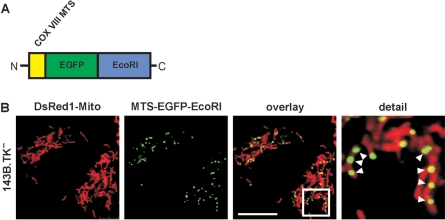
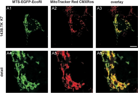
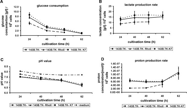
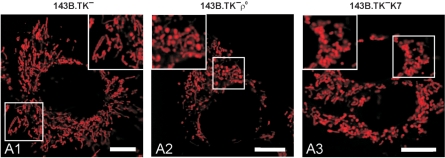
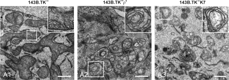
Eukaryotic cells devoid of mitochondrial DNA (rho0 cells) were originally generated under artificial growth conditions utilizing ethidium bromide. The chemical is known to intercalate preferentially with the mitochondrial double-stranded DNA thereby interfering with enzymes of the replication machinery. Rho0 cell lines are highly valuable tools to study human mitochondrial disorders because they can be utilized in cytoplasmic transfer experiments. However, mutagenic effects of ethidium bromide onto the nuclear DNA cannot be excluded. To foreclose this mutagenic character during the development of rho0 cell lines, we developed an extremely mild, reliable and timesaving method to generate rho0 cell lines within 3-5 days based on an enzymatic approach. Utilizing the genes for the restriction endonuclease EcoRI and the fluorescent protein EGFP that were fused to a mitochondrial targeting sequence, we developed a CMV-driven expression vector that allowed the temporal expression of the resulting fusion enzyme in eukaryotic cells. Applied on the human cell line 143B.TK- the active protein localized to mitochondria and induced the complete destruction of endogenous mtDNA. Mouse and rat rho0 cell lines were also successfully created with this approach. Furthermore, the newly established 143B.TK- rho0 cell line was characterized in great detail thereby releasing interesting insights into the morphology and ultra structure of human rho0 mitochondria.


Figures

Figure 1.
Figure 1.
Construction and expression of the mitochondrially targeted EcoRI. A DNA fragment coding for the COX VIII targeting sequence (MTS) was added to the 5′ end of the restriction endonuclease gene EcoRI and as optical marker the gene for the enhanced green fluorescent protein EGFP was fused to the 3′ end. The construct was sub-cloned into the vector pTRE2hyg and re-cloned with AgeI and NotI into pEGFP-Mito resulting in the vector pMEE-con with a constitutive CMV promoter (A) The final construct was transfected in 143B.DsRed1-Mito cells. Twenty-four hours after transfection the mitochondrial localization of the EcoRI fusion protein in punctate structures (white arrowheads) was confirmed by co-localization with DsRed1-Mito with confocal fluorescence microscopy (B) The calibration mark corresponds to 10 µm.
Figure 2.
Figure 2.
Graphic documentation of the proliferation rate of the cell lines 143B.TKK7 (dotted line), 143B.TK ρ0 (solid line) and 143B.TK (dashed line) at proceeding cultivation times. The indicated values are averages of four individual measurements. The standard deviations are depicted as error bars. The ρ0 cells exhibit a decelerated proliferation rate compared to the wild type. Time scale represents the time intervals proceeded after cell seeding.
Figure 3.
Figure 3.
Depletion of mtDNA in different cell lines after expression of a mitochondrially targeted EcoRI. (A) 143B.TK wild-type and 143B.TK ρ0 controls as well as the isolated 143B.TKK7 cells were analysed by PCR using primers corresponding to the D-loop region of mtDNA additionally to primers amplifying a histon H1 of nuclear DNA. It was not possible to amplify this mtDNA region in the cell lines 143B.TK ρ0 and 143B.TKK7 confirming the depletion of the mitochondrial genome in these cells. Positive amplification of histone H1 shows a sufficient amount of genomic DNA in all probes. (B) Southern blot analyses with these cells after digestion with BamHI or PvuII using a probe coding for mtDNA nucleotides 4831–5651 also showed the absence of mtDNA in the 143B.TK ρ0 and 143B.TKK7 cells (B). (C) LMTK (mouse) and NRK52E (rat) cells were treated and analysed as described above. mtDNA was undetectable in the newly established ρ0 cell lines of mouse and rat.
Figure 4.
Figure 4.
143B.TKK7 cells were transfected with pMEE-con and analysed by confocal fluorescence microscopy. The EGFP-tagged restriction enzyme (MTS-EGFP-EcoRI, green colour, A1 and A4) co-localizes (A3 and A6) with the MitoTracker Red CMXRos stained mitochondrial network (A2 and A5). When compared to Figure 1B, the punctate appearance (‘nucleoid’ structure) of MTS-EGFP-EcoRI merged into an even-stained mitochondrial network, indicating that the interacting partner (mtDNA) of the restriction enzyme (MTS-EGFP-EcoRI) disappeared thus underlining the ρ0 state of the cell.
Figure 5.
Figure 5.
Graphic documentation of the glucose consumption (A), lactate production rate (B) and proton production rate (C) in pure culture medium (dash-dotted line) and the culture medium of the cell lines 143B.TKK7 (dotted lines) 143B.TK ρ0 (solid lines) and 143B.TK (dashed lines) at proceeding cultivation times standardized to 106 cells. The indicated values are averages of four individual measurements, respectively eight independent measurements (pH value of ρ0 cells). The standard deviations are depicted as error bars. Time scale represents the time intervals proceeded after cell seeding. Standardized to 106 cells the glucose consumption as well as the lactate and proton production of the 143B.TKK7 and 143B.TK ρ0 cells exceed the amounts of the 143B.TK wild type. However, the pH value of culture medium in all determined cell lines was nearly similar. The pH value of pure medium without addition of cells shows a slight decline due to saturation of medium with CO2. An especial curve progression could be observed in the analysis of proton production rate where the values initially decline and then increase. It was not possible to determine the 62 h cultivation value of the wild-type cells because of extreme cell density resulting in apoptotic events.
Figure 6.
Figure 6.
Mitochondrial organization in wild-type and ρ0 143B.TK cells. The cells were stained with MitoTracker Red CMXRos. (A1) shows the predominantly reticular organization of mitochondria in wild-type cells with mainly rod-shaped organelles. 143B.TK ρ0 (A2) and 143B.TKK7 cells (A3) stained with MitoTracker Red CMXRos denotes the disruption of the mitochondrial reticulum in single units that often seemed to be swollen. Calibration marks correspond to 10 µm.
Figure 7.
Figure 7.
Ultra-structural investigation of wild-type and ρ0 mitochondria by TEM. Electron micrographs of ultra-thin sections are shown. 143B.TK wild-type mitochondria show an interconnected network structure with numerous regular arranged cristae (A1). The mitochondria of 143B.TK ρ0 (A2) and 143B.TKK7 cells (A3) demonstrate single vesicular organelles with distorted cristae, lying in concentric double membrane rings in the matrix. The insets show higher magnifications of the boxed areas. Bars (A1–A3), 1 µm.

References

    1. Wallace DC. A mitochondrial paradigm for degenerative diseases and ageing. Novartis Found. Symp. 2001;235:247–263. - PubMed
    1. Anderson S, Bankier AT, Barrell BG, de Bruijn MH, Coulson AR, Drouin J, Eperon IC, Nierlich DP, Roe BA, et al. Sequence and organization of the human mitochondrial genome. Nature. 1981;290:457–465. - PubMed
    1. King MP, Attardi G. Human cells lacking mtDNA: repopulation with exogenous mitochondria by complementation. Science. 1989;246:500–503. - PubMed
    1. Borst P, Grivell LA. The mitochondrial genome of yeast. Cell. 1978;15:705–723. - PubMed
    1. Goldring ES, Grossman LI, Krupnick D, Cryer DR, Marmur J. The petite mutation in yeast. Loss of mitochondrial deoxyribonucleic acid during induction of petites with ethidium bromide. J. Mol. Biol. 1970;52:323–335. - PubMed

Publication types

MeSH terms