. 2008 May;36(8):2705-16.
doi: 10.1093/nar/gkn102. Epub 2008 Mar 19.

Dissecting protein-RNA recognition sites

Affiliations

Dissecting protein-RNA recognition sites

Ranjit Prasad Bahadur et al. Nucleic Acids Res. 2008 May.

Abstract

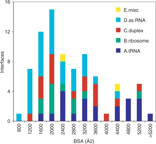
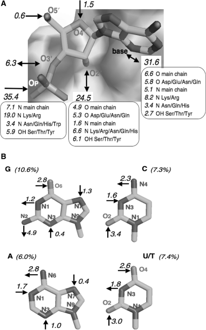
We analyze the protein-RNA interfaces in 81 transient binary complexes taken from the Protein Data Bank. Those with tRNA or duplex RNA are larger than with single-stranded RNA, and comparable in size to protein-DNA interfaces. The protein side bears a strong positive electrostatic potential and resembles protein-DNA interfaces in its amino acid composition. On the RNA side, the phosphate contributes less, and the sugar much more, to the interaction than in protein-DNA complexes. On average, protein-RNA interfaces contain 20 hydrogen bonds, 7 that involve the phosphates, 5 the sugar 2'OH, and 6 the bases, and 32 water molecules. The average H-bond density per unit buried surface area is less with tRNA or single-stranded RNA than with duplex RNA. The atomic packing is also less compact in interfaces with tRNA. On the protein side, the main chain NH and Arg/Lys side chains account for nearly half of all H-bonds to RNA; the main chain CO and side chain acceptor groups, for a quarter. The 2'OH is a major player in protein-RNA recognition, and shape complementarity an important determinant, whereas electrostatics and direct base-protein interactions play a lesser part than in protein-DNA recognition.


Figures

Figure 1.
Figure 1.
Size of protein–RNA interfaces. Histogram of the buried surface area (protein plus RNA) in each of the 81 complexes. The classes are defined in Table 1.
Figure 2.
Figure 2.
Buried interface area, atoms, residues and nucleotides. The number of interface atoms (A) and of interface amino acid residues or nucleotides (B) is plotted against the ASA lost by either the protein (x) or the RNA (•) component of the 81 complexes.
Figure 3.
Figure 3.
Shape and electrostatic potential of protein–RNA interfaces. The molecular surface of the proteins is colored according to its electrostatic potential; blue is positive and red negative. The RNA backbone is drawn as a tube. (A) The RNase E subunit binds a 15-mer RNA with the 5′-end at its active site (23); the interface is one of the smallest in our sample, but the 15-mer makes other contacts in the crystal (2bx2, class D). (B) The splicing endonuclease is a dimer (36); it forms an average size interface with a double-stranded 19-mer (2gjw, class C). (C) Yeast arginyl-tRNA synthetase (37) forms an extensive interface with tRNA-Arg (1f7u, class A). (D) Ribosomal protein S8 in complex with a 37-mer stem-loop fragment of 16S rRNA (38) (1i6u, class B). (E) The SAM domain of the Vts1 post-transcriptional regulator in complex with a 16-mer hairpin RNA (39) (2f8k, class D). (F) The 15.5 kDa spliceosomal protein in complex with a 22-mer stem-loop fragment of U4 snRNA (40) (1e7k, class C). The figure was created using PyMOL (DeLano Scientific LLC, San Carlos, CA, http://www.delanoscientific.com).
Figure 4.
Figure 4.
Euclidean distances between amino acid compositions. Values of Δf are calculated from the area based compositions in Table 4 as reported under ‘Results’ section.
Figure 5.
Figure 5.
The H-bonding pattern of RNA to proteins. The numbers are percent fractions of the 1637 protein–RNA H-bonds identified in the 81 complexes; a 5% fraction represents approximately one bond per complex. (A) Bonds involving the RNA backbone. (B) Bonds involving the bases. U/T includes pseudouracil.

References

    1. Berman HM, Battistuz T, Bhat TN, Bluhm WF, Bourne PE, Burkhardt K, et al. The Protein Data Bank. Acta Crystallog. Sect. 2002;D58:899–907. - PubMed
    1. Jones S, Thornton JM. Principles of protein-protein interactions. Proc. Natl Acad. Sci. USA. 1996;93:13–20. - PMC - PubMed
    1. Lo Conte L, Chothia C, Janin J. The atomic structure of protein–protein recognition sites. J. Mol. Biol. 1999;285:2177–2198. - PubMed
    1. Nadassy K, Wodak S, Janin J. Structural features of protein–nucleic acid recognition sites. Biochemistry. 1999;38:1999–2017. - PubMed
    1. Jones S, van Heyningen P, Berman HM, Thornton JM. Protein-DNA interactions: a structural analysis. J. Mol. Biol. 1999;287:877–896. - PubMed

Publication types