. 2008 May;294(5):C1169-74.
doi: 10.1152/ajpcell.00096.2008. Epub 2008 Mar 19.

Phosphorylation of beta-catenin by PKA promotes ATP-induced proliferation of vascular smooth muscle cells

Affiliations

Phosphorylation of beta-catenin by PKA promotes ATP-induced proliferation of vascular smooth muscle cells

Sebastien Taurin et al. Am J Physiol Cell Physiol. 2008 May.

Abstract

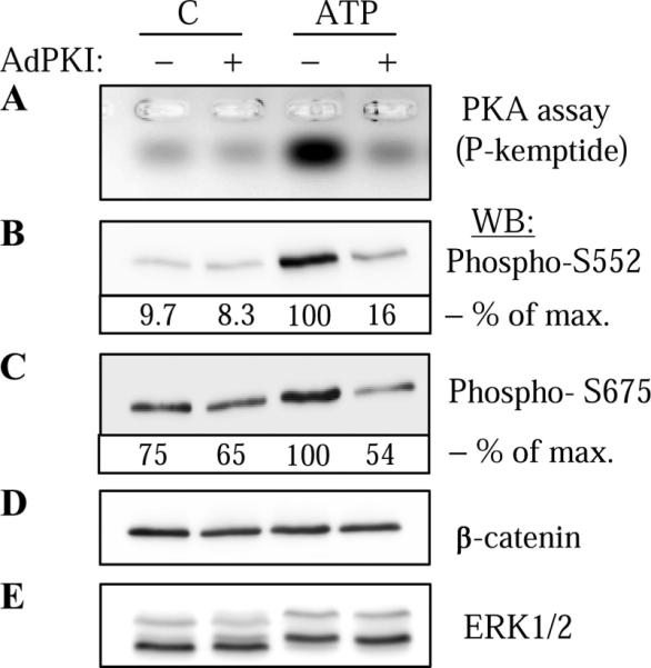
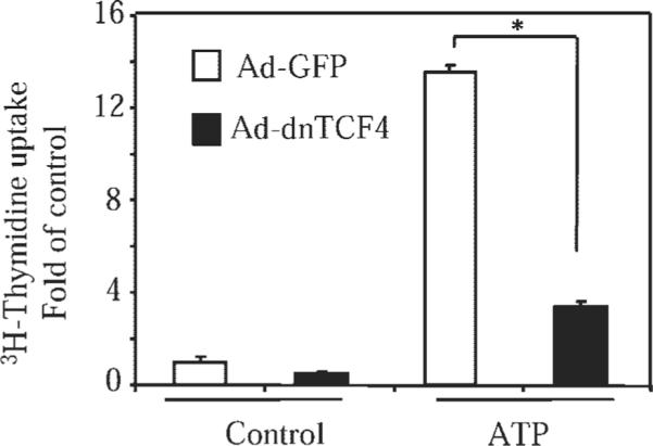
Extracellular ATP stimulates proliferation of vascular smooth muscle cells (VSMC) through activation of G protein-coupled P2Y purinergic receptors. We have previously shown that ATP stimulates a transient activation of protein kinase A (PKA), which, together with the established mitogenic signaling of purinergic receptors, promotes proliferation of VSMC (Hogarth DK, Sandbo N, Taurin S, Kolenko V, Miano JM, Dulin NO. Am J Physiol Cell Physiol 287: C449-C456, 2004). We also have shown that PKA can phosphorylate beta-catenin at two novel sites (Ser552 and Ser675) in vitro and in overexpression cell models (Taurin S, Sandbo N, Qin Y, Browning D, Dulin NO. J Biol Chem 281: 9971-9976, 2006). beta-Catenin promotes cell proliferation by activation of a family of T-cell factor (TCF) transcription factors, which drive the transcription of genes implicated in cell cycle progression including cyclin D1. In the present study, using the phosphospecific antibodies against phospho-Ser552 or phospho-Ser675 sites of beta-catenin, we show that ATP can stimulate PKA-dependent phosphorylation of endogenous beta-catenin at both of these sites without affecting its expression levels in VSMC. This translates to a PKA-dependent stimulation of TCF transcriptional activity through an increased association of phosphorylated (by PKA) beta-catenin with TCF-4. Using the PKA inhibitor PKI or dominant negative TCF-4 mutant, we show that ATP-induced cyclin D1 promoter activation, cyclin D1 protein expression, and proliferation of VSMC are all dependent on PKA and TCF activities. In conclusion, we show a novel mode of regulation of endogenous beta-catenin through its phosphorylation by PKA, and we demonstrate the importance of this mechanism for ATP-induced proliferation of VSMC.


Figures

Fig. 1
Fig. 1
Specificity of phospho-S552 and phospho-S675 β-catenin antibodies. Cos-7 cells were transfected with Flag-tagged β-catenin or the corresponding mutants of β-catenin and stimulated with 10 μM forskolin (FSK) for 5 min. The proteins were immunoprecipitated (IP) with Flag antibodies followed by Western blotting (WB) with phospho-S552 or phospho-S675 β-catenin antibodies as indicated. The equal amounts of immunoprecipitated β-catenin or the corresponding mutants were confirmed by Western blotting with Flag antibodies. WT, wild-type.
Fig. 2
Fig. 2
ATP-induced phosphorylation of endogenous β-catenin by PKA in vascular smooth muscle cells (VSMC). VSMC were transduced with control adenovirus (−) or adenovirus encoding PKA inhibitor PKI (Ad-PKI), stimulated with 30 μM ATP for 5 min and lysed. Cell lysates were analyzed for PKA activity (A) or were subjected to Western blotting with phospho-S552 (B) or phospho-S675 (C) β-catenin antibodies as indicated. The equal amounts of endogenous β-catenin were confirmed by Western blotting with β-catenin antibodies (D). For control purposes, ERK1/2 phosphorylation was assessed by electrophoretic mobility shift assay following Western blotting with ERK1/2 antibodies (E). The densitometry of selected blots is shown as % of maximal (max) response to ATP.
Fig. 3
Fig. 3
ATP-induced, PKA-mediated activation of T-cell factor (TCF)-dependent gene transcription in VSMC. A: PKA-dependent activation of TCF reporter by ATP. VSMC were transfected with cDNA for TCF-luciferase (Luc) reporter (TOPflash) or the mutated control reporter (FOPflash) along with a renilla reporter driven by thymidine kinase promoter (TK-RL), and with an empty vector or cDNA for PKA dominant negative mutant (dnPKA). Following the stimulation of cells with 30 μM ATP for 12 h, luciferase activity was measured and normalized to a corresponding renilla activity. The TOP-Luc/TK-RL values were subtracted from the FOP-Luc/TK-RL values. Data represent means ± SD from a representative of three experiments performed in triplicate. *P < 0.01. B: PKA-dependent interaction between endogenous TCF-4 and β-catenin in response to ATP. VSMC were transduced with Ad-PKI, stimulated with ATP for 5 min, and lysed. Endogenous TCF-4 was immunoprecipitated from cell lysates, and the immune complexes or total cell lysates were examined by Western blotting with desired antibodies as indicated. The densitometry of a selected blot is shown as % of maximal response to ATP.
Fig. 4
Fig. 4
ATP-induced activation of cyclin D1 promoter in VSMC is mediated by TCF-4 and PKA. VSMC were transfected with a control TK-RL DNA, a desired luciferase reporter for cyclin D1 promoter (A and D), TCF (Top/Fop) (B), or cAMP response element (CRE; C), along with the empty vector or the cDNAs for the TCF-4 dominant negative mutant (dnTCF-4; AC), or dnPKA (D). Following the stimulation of cells with 30 μM ATP for 12 h, luciferase activity was measured and normalized to a corresponding renilla activity. Data represent means ± SD from a representative of three experiments performed in triplicate. *P < 0.01.
Fig. 5
Fig. 5
ATP-induced activation of cyclin D1 protein expression in VSMC is mediated by TCF-4 and PKA. A: time course of cyclin D1 protein expression in response to 30 μM ATP as assessed by Western blotting with cyclin D1 antibodies. B and C: VSMC were transduced with control adenovirus or with Ad-PKI (B) or with Ad-dnTCF-4 (C). Cells were stimulated with 30 μM ATP for 12 h, and cell lysates were analyzed by Western blotting with antibodies against cyclin D1 and β-actin. The densitometry of selected blots is shown as % of maximal response to ATP.
Fig. 6
Fig. 6
ATP-induced DNA synthesis in VSMC is mediated by TCF-4. VSMC were transduced with control adenovirus [AD-green fluorescent protein (GFP)] or adenovirus encoding dnTCF-4 (Ad-dnTCF-4), followed by stimulation with 30 μM ATP for 12 h. The [3H]thymidine uptake assay was then performed as described in MATERIALS AND METHODS. Data represent means ± SD from a representative of three experiments performed in triplicate. *P < 0.01.

Similar articles

Cited by

References

    1. Abbott KL, Loss JR, 2nd, Robida AM, Murphy TJ. Evidence that Galpha(q)-coupled receptor-induced interleukin-6 mRNA in vascular smooth muscle cells involves the nuclear factor of activated T cells. Mol Pharmacol. 2000;58:946–953. - PubMed
    1. Aberle H, Bauer A, Stappert J, Kispert A, Kemler R. beta-Catenin is a target for the ubiquitin-proteasome pathway. EMBO J. 1997;16:3797–3804. - PMC - PubMed
    1. Abu-Amer Y, Ross FP, McHugh KP, Livolsi A, Peyron JF, Teitelbaum SL. Tumor necrosis factor-alpha activation of nuclear transcription factor-kappaB in marrow macrophages is mediated by c-Src tyrosine phosphorylation of Ikappa Balpha. J Biol Chem. 1998;273:29417–29423. - PubMed
    1. Alt JR, Cleveland JL, Hannink M, Diehl JA. Phosphorylation-dependent regulation of cyclin D1 nuclear export and cyclin D1-dependent cellular transformation. Genes Dev. 2000;14:3102–3114. - PMC - PubMed
    1. Bienz M, Clevers H. Linking colorectal cancer to Wnt signaling. Cell. 2000;103:311–320. - PubMed

Publication types

LinkOut - more resources