

State-dependent changes in glutamate, glycine, GABA, and dopamine levels in cat lumbar spinal cord
- PMID: 18353913
- PMCID: PMC2652136
- DOI: 10.1152/jn.01231.2007
State-dependent changes in glutamate, glycine, GABA, and dopamine levels in cat lumbar spinal cord
Abstract
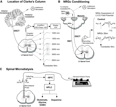
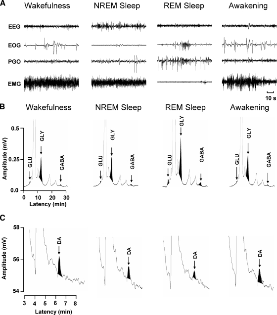
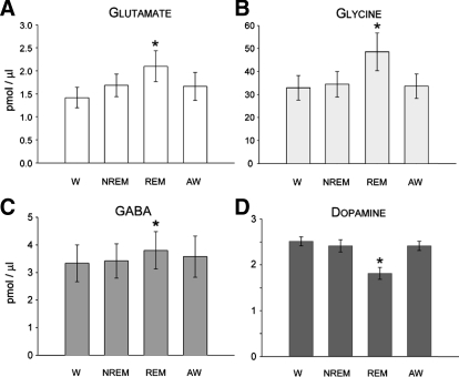
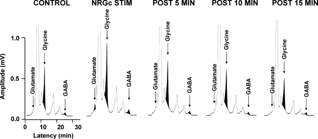
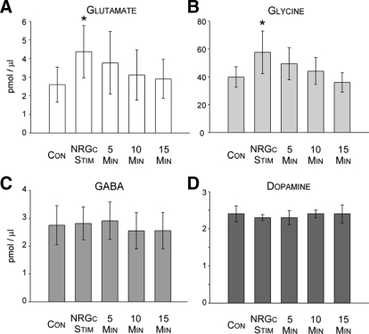
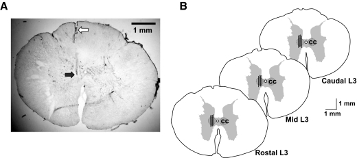
Recent studies have indicated that the glycine receptor antagonist strychnine and the gamma-aminobutyric acid type A (GABA A) receptor antagonist bicuculline reduced the rapid-eye-movement (REM) sleep-specific inhibition of sensory inflow via the dorsal spinocerebellar tract (DSCT). These findings imply that the spinal release of glycine and GABA may be due directly to the REM sleep-specific activation of reticulospinal neurons and/or glutamate-activated last-order spinal interneurons. This study used in vivo microdialysis and high-performance liquid chromatography analysis techniques to provide evidence for these possibilities. Microdialysis probes were stereotaxically positioned in the L3 spinal cord gray matter corresponding to sites where maximal cerebellar-evoked field potentials or individual DSCT and nearby spinoreticular tract (SRT) neurons could be recorded. Glutamate, glycine, and GABA levels significantly increased during REM sleep by approximately 48, 48, and 14%, respectively, compared with the control state of wakefulness. In contrast, dopamine levels significantly decreased by about 28% during REM sleep compared with wakefulness. During the state of wakefulness, electrical stimulation of the nucleus reticularis gigantocellularis (NRGc) at intensities sufficient to inhibit DSCT neuron activity, also significantly increased glutamate and glycine levels by about 69 and 45%, respectively, but not GABA or dopamine levels. We suggest that the reciprocal changes in the release of glutamate, glycine, and GABA versus dopamine during REM sleep contribute to the reduction of sensory inflow to higher brain centers via the DSCT and nearby SRT during this behavioral state. The neural pathways involved in this process likely include reticulo- and diencephalospinal and spinal interneurons.
Figures
Similar articles
-
On the reduction of spontaneous and glutamate-driven spinocerebellar and spinoreticular tract neuronal activity during active sleep.Neuroscience. 2001;104(1):199-206. doi: 10.1016/s0306-4522(01)00060-4. Neuroscience. 2001. PMID: 11311542
-
GABA release in the dorsal raphe nucleus: role in the control of REM sleep.Am J Physiol. 1997 Jul;273(1 Pt 2):R451-5. doi: 10.1152/ajpregu.1997.273.1.R451. Am J Physiol. 1997. PMID: 9249585 Free PMC article.
-
State-related inhibition by GABA and glycine of transmission in Clarke's column.J Neurosci. 2002 Jul 1;22(13):5777-88. doi: 10.1523/JNEUROSCI.22-13-05777.2002. J Neurosci. 2002. PMID: 12097531 Free PMC article.
-
[Selective stimulations and lesions of the rat brain nuclei as the models for research of the human sleep pathology mechanisms].Glas Srp Akad Nauka Med. 2011;(51):85-97. Glas Srp Akad Nauka Med. 2011. PMID: 22165729 Review. Serbian.
-
Neurophysiology of sleep and wakefulness.Respir Care Clin N Am. 2005 Dec;11(4):567-86. doi: 10.1016/j.rcc.2005.08.001. Respir Care Clin N Am. 2005. PMID: 16303589 Review.
Cited by
-
Behavioral response and transmitter release during atonia elicited by medial medullary stimulation.J Neurophysiol. 2010 Oct;104(4):2024-33. doi: 10.1152/jn.00528.2010. Epub 2010 Jul 28. J Neurophysiol. 2010. PMID: 20668280 Free PMC article.
-
Withania somnifera as an Adjunctive Treatment for Refractory Restless Legs Syndrome in Parkinson's Disease: A Case Report.Cureus. 2021 Dec 28;13(12):e20775. doi: 10.7759/cureus.20775. eCollection 2021 Dec. Cureus. 2021. PMID: 35111460 Free PMC article.
-
Sleep Deprivation and Recovery Sleep Prior to a Noxious Inflammatory Insult Influence Characteristics and Duration of Pain.Sleep. 2016 Jan 1;39(1):133-42. doi: 10.5665/sleep.5334. Sleep. 2016. PMID: 26237772 Free PMC article.
-
Rostro-caudal inhibition of hindlimb movements in the spinal cord of mice.PLoS One. 2014 Jun 25;9(6):e100865. doi: 10.1371/journal.pone.0100865. eCollection 2014. PLoS One. 2014. PMID: 24963653 Free PMC article.
-
Glycine-mediated postsynaptic inhibition is responsible for REM sleep atonia.Sleep. 2008 Nov;31(11):1483-6. doi: 10.1093/sleep/31.11.1483. Sleep. 2008. PMID: 19014067 Free PMC article. No abstract available.
References
-
- Bosco G, Poppele RE. Proprioception from a spinocerebellar perspective. Physiol Rev 81: 539–568, 2001. - PubMed
-
- Buchanan JT, Grillner S. Newly identified “glutamate interneurons” and their role in locomotion in the lamprey spinal cord. Science 236: 312–314, 1987. - PubMed
-
- Cechetto DF, Saper CB. Neurochemical organization of the hypothalamic projection to the spinal cord in the rat. J Comp Neurol 272: 579–604, 1988. - PubMed
-
- Chandler MJ, Garrison DW, Brennan TJ, Foreman RD. Effects of chemical and electrical stimulation of the midbrain on feline T2–T6 spinoreticular and spinal cell activity evoked by cardiopulmonary afferent input. Brain Res 496: 148–164, 1989. - PubMed
-
- Chase MH, Morales FR. The atonia and myoclonia of active (REM) sleep. Annu Rev Psychol 41: 557–584, 1990. - PubMed
Publication types
MeSH terms
Substances
Grants and funding
- R01 NS014610/NS/NINDS NIH HHS/United States
- HL-060296/HL/NHLBI NIH HHS/United States
- NS-14610/NS/NINDS NIH HHS/United States
- NS-34716/NS/NINDS NIH HHS/United States
- HL-41370/HL/NHLBI NIH HHS/United States
- R01 NS042566/NS/NINDS NIH HHS/United States
- P50 HL060296/HL/NHLBI NIH HHS/United States
- NS-32306/NS/NINDS NIH HHS/United States
- NS-041921/NS/NINDS NIH HHS/United States
- R01 HL041370/HL/NHLBI NIH HHS/United States
- R21 NS041921/NS/NINDS NIH HHS/United States
- R37 HL041370/HL/NHLBI NIH HHS/United States
- NS-042566/NS/NINDS NIH HHS/United States
- R37 NS014610/NS/NINDS NIH HHS/United States
LinkOut - more resources
Full Text Sources
Miscellaneous