. 2008 May;15(5):872-84.
doi: 10.1128/CVI.00463-07. Epub 2008 Mar 19.

Infection of macrophages and dendritic cells with primary R5-tropic human immunodeficiency virus type 1 inhibited by natural polyreactive anti-CCR5 antibodies purified from cervicovaginal secretions

Affiliations

Infection of macrophages and dendritic cells with primary R5-tropic human immunodeficiency virus type 1 inhibited by natural polyreactive anti-CCR5 antibodies purified from cervicovaginal secretions

Jobin Eslahpazir et al. Clin Vaccine Immunol. 2008 May.

Abstract

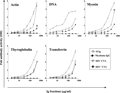
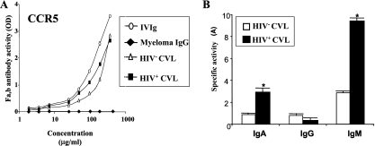
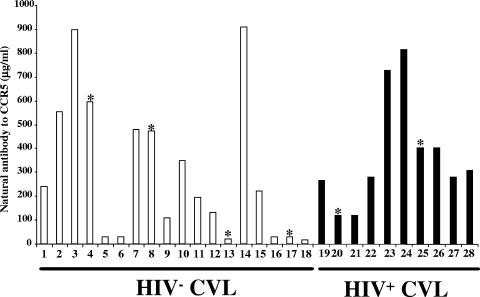
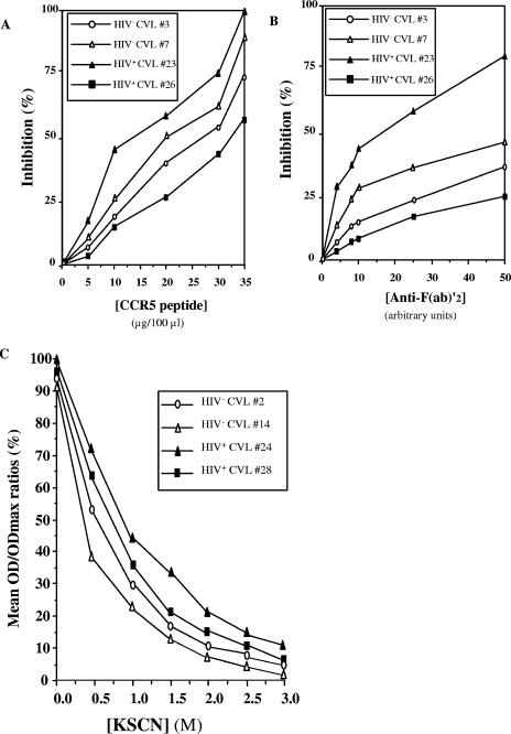
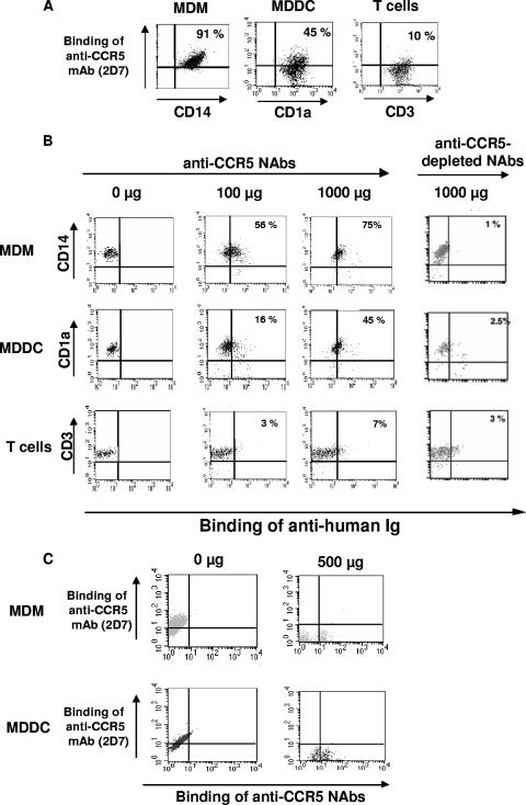
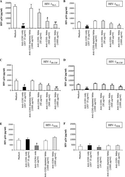
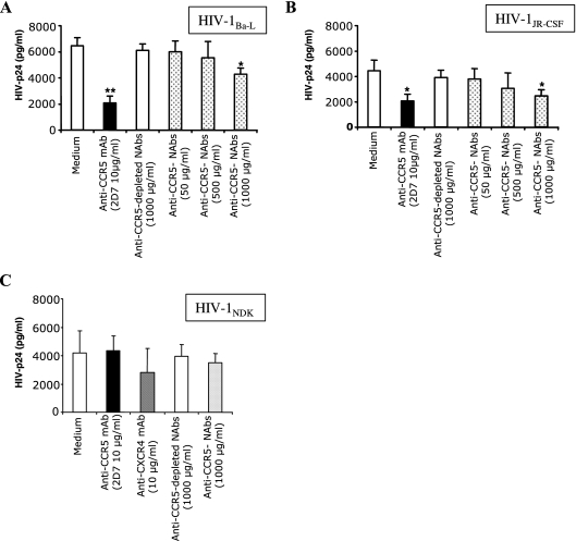
Heterosexual contact is the primary mode of human immunodeficiency virus (HIV) type 1 (HIV-1) transmission worldwide. The chemokine receptor CCR5 is the major coreceptor that is associated with the mucosal transmission of R5-tropic HIV-1 during sexual intercourse. The CCR5 molecule is thus a target for antibody-based therapeutic strategies aimed at blocking HIV-1 entry into cells. We have previously demonstrated that polyreactive natural antibodies (NAbs) from therapeutic preparations of immunoglobulin G and from human breast milk contain NAbs directed against CCR5. Such antibodies inhibit the infection of human macrophages and T lymphocytes by R5-tropic isolates of HIV in vitro. In the present study, we demonstrate that human immunoglobulins from the cervicovaginal secretions of HIV-seronegative or HIV-seropositive women contain NAbs directed against the HIV-1 coreceptor CCR5. Natural affinity-purified anti-CCR5 antibodies bound to CCR5 expressed on macrophages and dendritic cells and further inhibited the infection of macrophages and dendritic cells with primary and laboratory-adapted R5-tropic HIV but not with X4-tropic HIV. Natural anti-CCR5 antibodies moderately inhibited R5-tropic HIV transfer from monocyte-derived dendritic cells to autologous T cells. Our results suggest that mucosal anti-CCR5 antibodies from healthy immunocompetent donors may hamper the penetration of HIV and may be suitable for use in the development of novel passive immunotherapy regimens in specific clinical settings of HIV infection.


Figures

FIG. 1.
FIG. 1.
Autoantibody reactivities to bovine actin, double-stranded human DNA, porcine myosin, porcine thyroglobulin, and human transferrin in total Ig fractions purified from pooled cervicovaginal secretions. Antigens were coated on 96-well plates; and serial dilutions of i.v. Ig (open circles), IgG myeloma (bold diamonds), or pooled cervicovaginal secretions from HIV-seronegative women (HIV; open triangles) or HIV-seropositive women (HIV+; bold squares) were further added, as described in Materials and Methods. The concentrations of the antigen-reacting antibodies were evaluated by the addition of biotin-conjugated mouse Ig against anti-human Fab, followed by the addition of streptavidin-peroxidase, and the results were revealed by substrate addition. The total Ig content (IgA, IgG, and IgM) of the preparations is shown on the abscissa. The results are expressed as the means of the values obtained from three independent experiments ± SDs. The reactivity intensities for actin and myosin between pools from HIV-seronegative and HIV-seropositive women were significantly different (P < 0.01) only for high antibody concentrations (500 μg/ml); *, P ≤ 0.01 by the paired Student t test between the specific activities of CVL fluid samples from HIV-seronegative and HIV-seropositive women.
FIG. 2.
FIG. 2.
Autoantibody reactivities to CCR5. (A) CCR5 peptide CSSHFPYSQYQFWKNFQTLK, which corresponds to the second extracellular loop of CCR5 (II.E/C-CCR5), was coated on a 96-well plate; and serial dilution of i.v. Ig (open circles), IgG myeloma (bold diamonds), or pooled cervicovaginal secretions from HIV-seronegative women (HIV; open triangles) or HIV-seropositive women (HIV+; bold square) were further added, as described in Materials and Methods. The concentration of CCR5-reacting antibodies was evaluated by the addition of biotin-conjugated mouse Ig against anti-human Fab, followed by the addition of streptavidin-peroxidase, and the results were revealed by substrate addition. The total Ig content (IgA, IgG, and IgM) of the preparations is shown on the abscissa. The results are expressed as the means of the values obtained from three independent experiments ± SDs. (B). Specific activities, in arbitrary units of IgA, IgG, and IgM NAbs, to CCR5 in pools of cervicovaginal secretions from HIV-seronegative healthy women (open squares) or HIV-seropositive women (bold squares) by isotype. The specific activities of antibodies of the IgA, IgG, or IgM isotype to the CCR5 peptide in CVL fluid samples were evaluated at the first dilution that gave an OD between 0.5 and 1.5 and per μg of total IgA, IgG, and IgM antibodies in CVL fluid samples, respectively, according to the formula A = (OD × d)/[Ig] (in μg/ml), as described previously (3, 40). The results are expressed as the means of the values obtained from three independent experiments ± SDs. *, P ≤ 0.05 by the paired Student t test between specific activities of CVL fluid samples from HIV-seronegative and HIV-seropositive women.
FIG. 3.
FIG. 3.
Reactivities toward the CCR5 peptide in cervicovaginal secretion samples from 18 HIV-seronegative women (HIV) and 10 HIV-1-infected women (HIV+). Autoantibody activities toward the CCR5 peptide were detected as described in Materials and Methods. Each point represents the mean OD of duplicate assays. The results are expressed in μg/ml. According to the threshold of the indirect ELISA used (50 μg/ml), all CVL fluid samples except those from women 5, 6, 13, 16, 17, and 18 contained relevant amounts of CCR5-specific antibodies. *, women whose cervicovaginal secretion samples contained traces of semen.
FIG. 4.
FIG. 4.
(A and B) Specificities of anti-CCR5 NAbs. The CCR5 peptide was coated on a 96-well plate, and anti-CCR5 NAbs purified from CVL fluid samples from HIV-seronegative women (HIV; samples 3 and 7) and HIV-seropositive women (HIV+; samples 23 and 26) were further added in the presence of increasing amounts of CCR5 peptide (A) and anti-F(ab′)2 antibodies (B), as described in Materials and Methods. The reactivities to the coated CCR5 peptide were detected as described in Materials and Methods. The results, obtained from three independent experiments, are expressed as the mean percent inhibition of binding of NAbs to coated-CCR5 peptide ± SD. (C) Avidities of anti-CCR5 NAbs. Anti-CCR5 antibodies purified from CVL fluid samples from HIV-seronegative women (samples 2 and 14) and HIV-seropositive women (samples 24 and 28) were incubated with the CCR5 peptide for 2 h at 37°C before the wells were treated with increasing amounts of KSCN (0.1 M to 3 M) for 30 min at room temperature. The wells were washed and incubated with goat anti-human F(ab′)2 coupled to peroxidase for 1 h at 37°C. The molarity of KSCN required for the dissociation of 50% of the bound antibodies was then determined. The avidity index was defined as the molarity of KSCN equivalent to the interpolation point corresponding to 50% of the control absorbance value obtained in the absence of KSCN.
FIG. 5.
FIG. 5.
Binding of anti-CCR5 NAbs to macrophages (CD14+), immature dendritic cells (CD1a+), and T lymphocytes (CD3+) at the single-cell level. (A) MDMs, MDDCs, and T lymphocytes (105) were incubated with anti-CD14-FITC, anti-CD1a-FITC, and anti-CD3-FITC MAbs, respectively, and with an anti-CCR5 PE MAb (2D5 clone; 10 μg/ml) at 4°C for 30 min. (B) MDMs, MDDCs, and T lymphocytes (105) were incubated with mouse anti-CD14 PE, anti-CD1a PE, and anti-CD3-PE MAbs, respectively, and with either 0, 100, or 1,000 μg/ml of purified anti-CCR5 NAbs or 1,000 μg/ml of anti-CCR5-depleted NAbs at 4°C for 30 min. The cells were subsequently incubated with FITC-conjugated goat anti-human F(ab′)2 antibody at 4°C for 30 min. (C) MDMs and MDDCs (105) were incubated with or without 1,000 μg/ml of purified anti-CCR5 NAbs at 4°C for 30 min. The cells were subsequently incubated with FITC-conjugated goat anti-human F(ab′)2 antibody and an anti-CCR5 PE MAb at 4°C for 30 min. The percentage of positive cells is indicated in quadrants defined according to the relevant isotypic control. The results of one representative experiment of four independent experiments conducted are shown.
FIG. 6.
FIG. 6.
Inhibition of infection of macrophages and dendritic cells with R5-HIVBaL and R5-HIVJR-CSF by anti-CCR5 NAbs purified from CVL fluid samples from HIV-seronegative women. MDMs (A, C, and E) and MDDCs (B, D, and F) were preincubated with either anti-CCR5 affinity-purified NAbs (50, 500, and 1,000 μg/ml), anti-CCR5-depleted NAbs (1,000 μg/ml; negative control), anti-CCR5 MAb 2D7 (10 μg/ml), or an anti-CXCR4 MAb (10 μg/ml) prior to infection with R5-HIVBaL (A and B), R5-HIVJR-CSF (C and D), or X4-HIVNDK (1 ng/ml) (E and F) at 37°C for 3 h. The level of production of the HIV p24 antigen in the culture supernatants was determined at day 6 postinfection by ELISA. The results are expressed as the mean HIV p24 concentrations from three independent experiments ± SDs. *, P ≤ 0.05 by the paired Student t test compared to the HIV p24 concentration in medium; **, P ≤ 0.01 by the paired Student t test compared to the HIV p24 concentration in medium.
FIG. 7.
FIG. 7.
Partial inhibition of R5-HIVBaL and R5-HIVJR-CSF transfer to T cells from MDDCs by anti-CCR5 NAbs purified from CVL fluid samples from HIV-seronegative women. MDDCs were preincubated with either anti-CCR5 affinity-purified NAbs (50, 500, and 1,000 μg/ml), anti-CCR5-depleted NAbs (1,000 μg/ml; negative control), anti-CCR5 MAb 2D7 (10 μg/ml), or an anti-CXCR4 MAb (10 μg/ml) prior to infection with R5-HIVBaL (A), R5-HIVJR-CSF (B), or X4-HIVNDK (C) (1 ng/ml) at 37°C for 3 h. Following extensive washes, autologous activated T cells (see Materials and Methods) were cocultured with infected MDDCs at a dendritic cell-to-T-cell ratio of 1:5. The production of the HIV p24 antigen in the culture supernatants was determined at day 3 postinfection by ELISA. The results are expressed as the mean HIV p24 concentrations from three independent experiments ± SDs. *, P ≤ 0.05 by the paired Student t test compared to the HIV p24 concentration in medium; **, P ≤ 0.01 by the paired Student t test compared to the HIV p24 concentration in medium.

Similar articles

Cited by

References

    1. Avrameas, S., G. Dighiero, P. Lymberi, and B. Guilbert. 1983. Studies on natural antibodies and autoantibodies. Ann. Immunol. (Paris) 134D:103-113. - PubMed
    1. Bard, E., D. Riethmuller, S. Biichle, D. Meillet, J. L. Pretet, C. Mougin, and E. Seilles. 2002. Validation of a high sensitive immunoenzymatic assay to establish the origin of immunoglobulins in female genital secretions. J. Immunoassay Immunochem. 23:145-162. - PubMed
    1. Baumgarth, N., J. W. Tung, and L. A. Herzenberg. 2005. Inherent specificities in natural antibodies: a key to immune defense against pathogen invasion. Springer Semin. Immunopathol. 26:347-362. - PubMed
    1. Belec, L. 2002. Defenses of the female genital tract against infection. J. Gynecol. Obstet. Biol. Reprod (Paris) 31:4S45-4S59. (In French.) - PubMed
    1. Belec, L., T. Dupre, T. Prazuck, C. Tevi-Benissan, J. M. Kanga, O. Pathey, X. S. Lu, and J. Pillot. 1995. Cervicovaginal overproduction of specific IgG to human immunodeficiency virus (HIV) contrasts with normal or impaired IgA local response in HIV infection. J. Infect. Dis. 172:691-697. - PubMed

Publication types

LinkOut - more resources