. 2008 Jun;82(11):5153-60.
doi: 10.1128/JVI.00162-08. Epub 2008 Mar 19.

Vaccinia virus A56/K2 fusion regulatory protein interacts with the A16 and G9 subunits of the entry fusion complex

Affiliations

Vaccinia virus A56/K2 fusion regulatory protein interacts with the A16 and G9 subunits of the entry fusion complex

Timothy R Wagenaar et al. J Virol. 2008 Jun.

Abstract

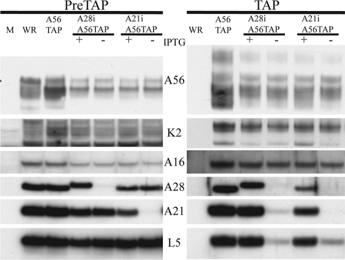
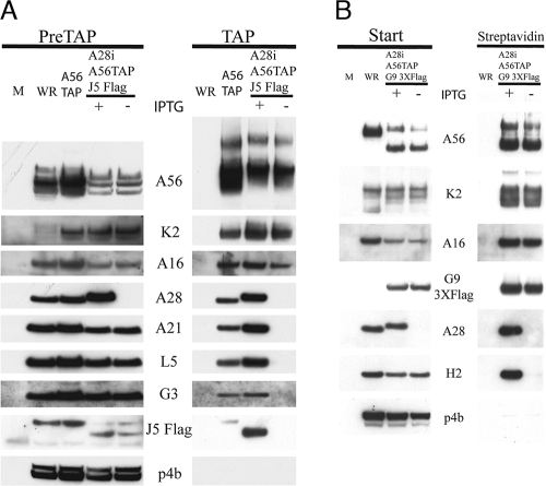
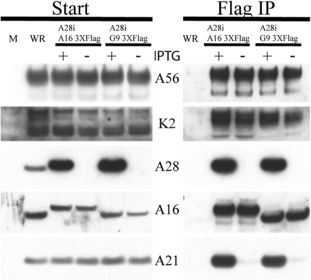
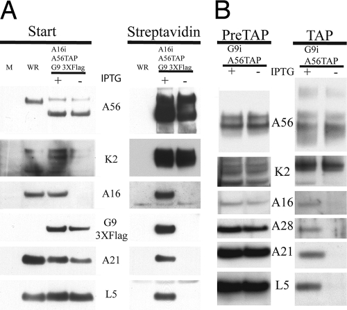
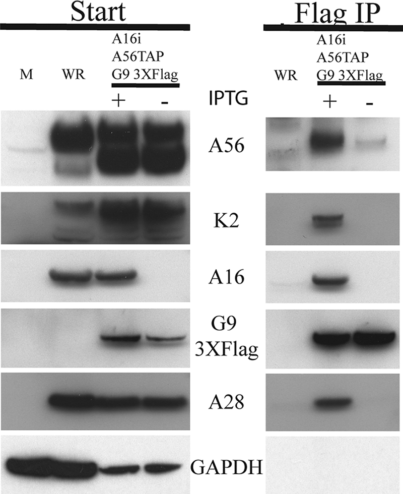
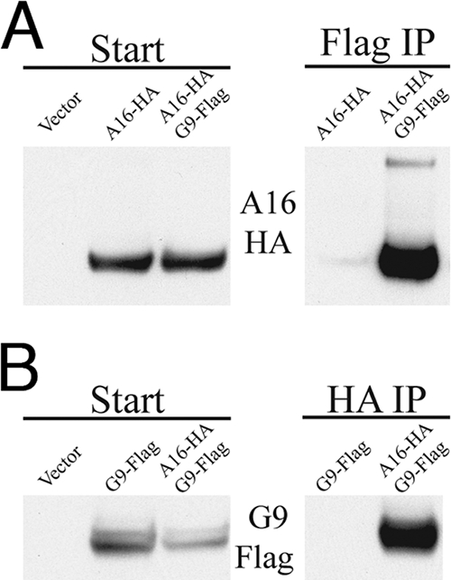
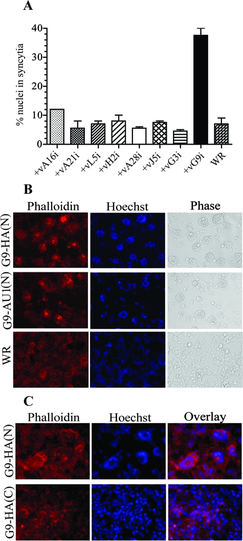
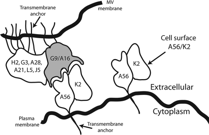
Deletion of the A56R or K2L gene of vaccinia virus (VACV) results in the spontaneous fusion of infected cells to form large multinucleated syncytia. A56 and K2 polypeptides bind to one another (A56/K2) and together are required for interaction with the VACV entry fusion complex (EFC); this association has been proposed to prevent the fusion of infected cells. At least eight viral polypeptides comprise the EFC, but no information has been available regarding their interactions either with each other or with A56/K2. Utilizing a panel of recombinant VACVs designed to repress expression of individual EFC subunits, we demonstrated that A56/K2 interacted with two polypeptides: A16 and G9. Both A16 and G9 were required for the efficient binding of each to A56/K2, suggesting that the two polypeptides interact with each other within the EFC. Such an interaction was established by the copurification of A16 and G9 from infected cells under conditions in which a stable EFC complex failed to assemble and from detergent-treated lysates of uninfected cells that coexpressed A16 and G9. A recombinant VACV that expressed G9 modified with an N-terminal epitope tag induced the formation of syncytia, suggesting partial interference with the functional interaction of A56/K2 with the EFC during infection. These data suggest that A16 and G9 are physically associated within the EFC and that their interaction with A56/K2 suppresses spontaneous syncytium formation and possibly "fuse-back" superinfection of cells.


Figures

FIG. 1.
FIG. 1.
A56/K2 physically associates with A16. HeLa cells were mock infected (M) or infected with VACV WR, vA56TAP, vA28iA56TAP (with [+] or without [−] IPTG), or vA21iA56TAP (with or without IPTG). After 24 h, the cells were lysed with Triton X-100 and the A56 protein was tandem affinity purified. The starting material (PreTAP) and purified samples (TAP) were separated by SDS-PAGE and analyzed by Western blotting, using antibodies to the entry proteins A16, A28, A21, and L5, as well as antibodies to the A56 and K2 proteins. Secondary antibodies conjugated to horseradish peroxidase were used for detection by chemiluminescence. The recombinant viruses are listed at the top, and the targets of the specific antibodies are listed on the side.
FIG. 2.
FIG. 2.
A16 and G9 selectively copurify with A56/K2. (A) HeLa cells were mock infected (M) or infected with VACV WR, vA56TAP, or vA28iA56TAPJ5Flag with (+) or without (−) IPTG. After 24 h, the A56 protein was isolated from the infected cell lysates by successive bindings to streptavidin and calmodulin beads. Western blotting was performed on the starting material (PreTAP) and affinity purified proteins (TAP) as described in the legend to Fig. 1. (B) BS-C-1 cells were mock infected or infected with VACV WR or vA28iA56TAPG93XFlag (with or without IPTG). After 24 h, the A56 proteins were isolated by binding to streptavidin Sepharose. Western blotting was performed on the starting material (Start) and affinity purified proteins (Streptavidin) as described in the legend to Fig. 1.
FIG. 3.
FIG. 3.
A56/K2 binds epitope-tagged A16 and G9. BS-C-1 cells were mock infected (M) or infected with VACV WR, vA28iA163XFlag (with [+] or without [−] IPTG), or vA28iG93XFlag (with or without IPTG). Infected cells were harvested after 24 h and lysed with Triton X-100. The lysate was cleared by centrifugation, and the A16 and G9 polypeptides were isolated by binding to agarose beads conjugated with Flag antibody. The eluate (Flag IP) and starting material (Start) were separated by SDS-PAGE and analyzed by Western blotting with antibodies to the viral proteins A56, K2, A28, A16, and A21 as indicated on the side. The viruses are indicated at the top of the figure.
FIG. 4.
FIG. 4.
Both A16 and G9 are required for binding A56/K2. (A) BS-C-1 cells were mock infected (M) or infected with VACV WR or vA16iA56TAPG93XFlag (with [+] or without [−] IPTG). The cells were lysed with Triton X-100 after 24 h, and the A56 protein was isolated by binding to streptavidin beads. The starting material (Start) and the affinity-purified proteins (Streptavidin) were resolved by SDS-PAGE and analyzed by Western blotting, using antibodies to the viral proteins indicated on the side. (B) HeLa cells were infected with vG9iA56TAP (with or without IPTG), and the A56 proteins were tandem affinity purified. The starting material (PreTAP) and the purified proteins (TAP) were analyzed as for panel A.
FIG. 5.
FIG. 5.
A16 is required for G9 to bind A56/K2. Cells were mock infected (M) or infected with VACV WR or vA16iA56TAPG93XFlag (with [+] or without[−] IPTG). Infected BS-C-1 cells were harvested at 24 h and lysed with Triton X-100. Flag antibody conjugated to agarose beads was used to purify the G9 protein. The bound material (Flag IP) was eluted from the agarose beads and separated along with the starting material (Start) by SDS-PAGE and then transferred to nitrocellulose. Western blotting was performed, using antibodies to the proteins A56, K2, A16, or A28, the Flag epitope, or glyceraldehyde phosphate dehydrogenase (GAPDH), as indicated.
FIG. 6.
FIG. 6.
A16 and G9 interact in uninfected cells. (A) 293TT cells were transfected with empty vector or plasmid DNA expressing A16 with a C-terminal influenza virus HA epitope (A16-HA) or cotransfected with plasmids expressing A16-HA and G9 with a C-terminal 3XFlag tag (G9-Flag). After 48 h, the cells were lysed with Triton X-100, the postnuclear supernatant was incubated with the Flag antibody bound to beads. The starting material (Start) and the captured proteins (Flag IP) were analyzed by Western blotting with the anti-HA antibody. (B) Cells were transfected with empty vector or G9-Flag or cotransfected with A16-HA and G9-3XFlag. The lysates were incubated with anti-HA antibody bound to beads. The starting material (Start) and the captured proteins (HA IP) were analyzed by Western blotting with the anti-Flag antibody.
FIG. 7.
FIG. 7.
Syncytium formation induced by modified G9. (A) HeLa cell monolayers were infected with 2 PFU per cell of VACVs expressing inducible A16, A21, L5, H2, A28, J5, G3, G9, or VACV WR for 1 h at 37°C and then washed and incubated for 21 h in the presence of IPTG. The cells were stained with Hoechst dye to visualize DNA and Alexa Fluor 594-phalloidin to visualize the actin cytoskeleton. The percentage of nuclei in syncytia was determined by counting the number of nuclei in fused cells that contained three or more nuclei and dividing by the total number of nuclei. Standard error bars are shown. (B and C) HeLa cell monolayers were infected with 2 PFU per cell of VACV expressing G9 with an HA epitope tag at the N or C terminus or an AU1 tag at the N terminus for 21 h and then stained as for panel A. Fluorescent and phase-contrast microscopic images are shown.
FIG. 8.
FIG. 8.
Model of G9/A16 in MV membrane binding to A56/K2 in plasma membrane. G9 and A16 are anchored in MV membrane in association with the EFC. A56/K2 is anchored in plasma membrane through the transmembrane domain of A56. The interaction of A56/K2 with A16 and G9 is postulated to prevent fusion of the MV particle with the plasma membrane.

References

    1. Armstrong, J. A., D. H. Metz, and M. R. Young. 1973. The mode of entry of vaccinia virus into L cells. J. Gen. Virol. 21533-537. - PubMed
    1. Blasco, R., and B. Moss. 1992. Role of cell-associated enveloped vaccinia virus in cell-to-cell spread. J. Virol. 664170-4179. - PMC - PubMed
    1. Brown, C. K., P. C. Turner, and R. W. Moyer. 1991. Molecular characterization of the vaccinia virus hemagglutinin gene. J. Virol. 653598-3606. - PMC - PubMed
    1. Brown, E., T. G. Senkevich, and B. Moss. 2006. Vaccinia virus F9 virion membrane protein is required for entry but not virus assembly, in contrast to the related L1 protein. J. Virol. 809455-9464. - PMC - PubMed
    1. Brum, L. M., P. C. Turner, H. Devick, M. T. Baquero, and R. W. Moyer. 2003. Plasma membrane localization and fusion inhibitory activity of the cowpox virus serpin SPI-3 require a functional signal sequence and the virus encoded hemagglutinin. Virology 306289-302. - PubMed

Publication types

LinkOut - more resources