. 2008 Apr 1;105(13):5219-24.
doi: 10.1073/pnas.0710301105. Epub 2008 Mar 19.

Structural basis of GM1 ganglioside recognition by simian virus 40

Affiliations

Structural basis of GM1 ganglioside recognition by simian virus 40

Ursula Neu et al. Proc Natl Acad Sci U S A. .

Abstract

Simian virus 40 (SV40) has been a paradigm for understanding attachment and entry of nonenveloped viruses, viral DNA replication, and virus assembly, as well as for endocytosis pathways associated with caveolin and cholesterol. We find by glycan array screening that SV40 recognizes its ganglioside receptor GM1 with a quite narrow specificity, but isothermal titration calorimetry shows that individual binding sites have a relatively low affinity, with a millimolar dissociation constant. The high-resolution crystal structure of recombinantly produced SV40 capsid protein, VP1, in complex with the carbohydrate portion of GM1, reveals that the receptor is bound in a shallow solvent-exposed groove at the outer surface of the capsid. Through a complex network of interactions, VP1 recognizes a conformation of GM1 that is the dominant one in solution. Analysis of contacts provides a structural basis for the observed specificity and suggests binding mechanisms for additional physiologically relevant GM1 variants. Comparison with murine Polyomavirus (Polyoma) receptor complexes reveals that SV40 uses a different mechanism of sialic acid binding, which has implications for receptor binding of human polyomaviruses. The SV40-GM1 complex reveals a parallel to cholera toxin, which uses a similar cell entry pathway and binds GM1 in the same conformation.


Conflict of interest statement

The authors declare no conflict of interest.

Figures
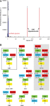
Fig. 1.
Fig. 1.
Specificity of SV40 VP1 for GM1. (A) Analysis of specificity by glycan array screening. Glycan array data are given as mean fluorescence signal for each glycan (see Materials and Methods). Error bars correspond to the standard error of the mean of six replicates for each glycan, with the highest and lowest signals omitted from analysis to reduce bias from extreme values. The signals on the array either come from glycoproteins (blue) or from synthesized carbohydrates (red). (B) Schematic view of gangliosides and related structures present on the array. GM1 is highlighted with a black box, and selected gangliosides not included in the array but discussed in the text are shaded gray.
Fig. 2.
Fig. 2.
Structure of the SV40 VP1–GM1 complex. (A) Overvall structure. The five VP1 chains are shown as ribbon tracings, with one VP1 monomer highlighted in blue and the others colored gray. Bound GM1 is shown in stick representation, with carbons drawn in orange, oxygens in red and nitrogens in blue. The GM1 molecules bind on top of the VP1 pentamer, corresponding to the capsid surface. (B) Composite annealed omit difference electron density map for GM1, calculated at 2.25-Å resolution, contoured at 2.5 σ and displayed 4 Å around GM1. One VP1 monomer is colored blue, the clockwise (cw) and counterclockwise (ccw) neighbors are colored gray.
Fig. 3.
Fig. 3.
Interactions between SV40 VP1 and GM1. (A) Surface architecture of VP1 and shape complementarity with GM1. Protein chains are colored using the code in Fig. 2. (B) Contacts between NeuNAc and SV40 VP1. (C) Contacts between Gal-(β1,3)-GalNAc and SV40 VP1. In B and C, only residues that contact the sugar are shown in stick representation. Hydrogen bonds are shown as broken lines. The cavity between one monomer of VP1 and its clockwise neighbor is shaded gray.
Fig. 4.
Fig. 4.
SV40 and Polyoma bind their ganglioside receptors in different orientations and conformations. (A and B) Comparison of specific interactions between VP1 from SV40 (A) and Polyoma (B) with their ligands. Polyoma was crystallized with an oligosaccharide that contained a fragment also present in GD1a and GT1b (shown in color). The additional α2,6-linked NeuNAc present in the ligand, but not on a ganglioside receptor, is shown in gray. The same view was used for both proteins, and residues at equivalent positions in both proteins have the same color. The Arg-Gly motif of Polyoma and its SV40 counterpart are colored yellow, and the residues holding them in place are shown in green. Hydrophobic residues lining the deep cavity in SV40 and their charged Polyoma equivalents are turquoise. Additional residues contacting NeuNAc in both complexes are gray. (C and D) Different binding surfaces of SV40 (C) and Polyoma VP1 (D) for their ligands. The same view from top of the pentamer was used for both complexes, and the color scheme is as in A and B. The terminal Glc was omitted from the SV40 VP1-GM1 complex for clarity. (E) Alignment of the VP1 sequences of SV40, Polyoma, BKV, and JCV. A sequence alignment of SV40, BKV, and JCV VP1 was performed with T-Coffee (www.ebi.ac.uk/t-coffee), then the aligned sequences were arranged according to a structure-based alignment of SV40 and Polyoma (18). SV40 residues identical to both BKV and JCV are marked with an asterisk; those identical to either BKV or JCV are marked with a dot. The color scheme is as in A and B, with additional SV40 residues in contact with GM1 in gray.

Comment in

Similar articles

Cited by

References

    1. Eddy BE, Borman GS, Grubbs GE, Young RD. Identification of the oncogenic substance in rhesus monkey kidney cell culture as simian virus 40. Virology. 1962;17:65–75. - PubMed
    1. Girardi AJ, Sweet BH, Slotnick VB, Hilleman MR. Development of tumors in hamsters inoculated in the neonatal period with vacuolating virus, SV-40. Proc Soc Exp Biol Med. 1962;109:649–660. - PubMed
    1. Berger JR, Major EO. Progressive multifocal leukoencephalopathy. Semin Neurol. 1999;19:193–200. - PubMed
    1. Drachenberg CB, et al. Human polyoma virus in renal allograft biopsies: morphological findings and correlation with urine cytology. Hum Pathol. 1999;30:970–977. - PubMed
    1. Stehle T, Gamblin SJ, Yan Y, Harrison SC. The structure of simian virus 40 refined at 3.1 Å resolution. Structure (London) 1996;4:165–182. - PubMed

Publication types

LinkOut - more resources