. 2008 Mar 25;105(12):4615-20.
doi: 10.1073/pnas.0708058105. Epub 2008 Mar 19.

A two-step nucleotide-flipping mechanism enables kinetic discrimination of DNA lesions by AGT

Affiliations

A two-step nucleotide-flipping mechanism enables kinetic discrimination of DNA lesions by AGT

Jie Hu et al. Proc Natl Acad Sci U S A. .

Abstract

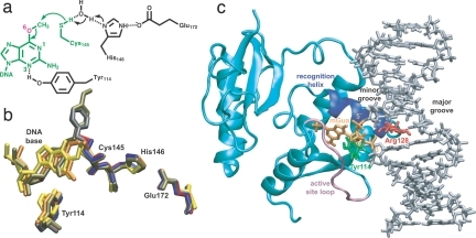
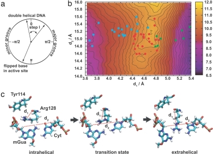
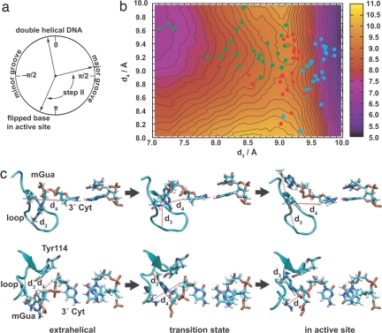
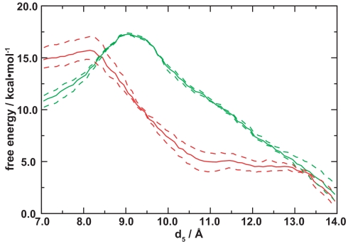
O(6)-alkylguanine-DNA alkyltransferase (AGT) repairs damage to the human genome by flipping guanine and thymine bases into its active site for irreversible transfer of alkyl lesions to Cys-145, but how the protein identifies its targets has remained unknown. Understanding molecular recognition in this system, which can serve as a paradigm for the many nucleotide-flipping proteins that regulate genes and repair DNA in all kingdoms of life, is particularly important given that inhibitors are in clinical trials as anticancer therapeutics. Computational approaches introduced recently for harvesting and statistically characterizing trajectories of molecularly rare events now enable us to elucidate a pathway for nucleotide flipping by AGT and the forces that promote it. In contrast to previously proposed flipping mechanisms, we observe a two-step process that promotes a kinetic, rather than a thermodynamic, gate-keeping strategy for lesion discrimination. Connection is made to recent single-molecule studies of DNA-repair proteins sliding on DNA to understand how they sense subtle chemical differences between bases efficiently.


Conflict of interest statement

The authors declare no conflict of interest.

Figures

Fig. 1.
Fig. 1.
Structure of AGT. (a) Chemical mechanism (21). (b) Comparison of active-site structures: blue, unmodified wt (PDB entry 1EH6) (17); red, wt with methylated Cys-145 (1EH7) (17); gray, wt with benzylated Cys-145 (1EH8) (17); orange, C145S mutant with DNA containing mGua (1T38) (21); yellow, wt with DNA containing N1,O6-ethanoxanthine (1T39) (21); tan, wt bound to N4-alkylcytosine (1YFH structure C) (22). (c) Wild-type–mGua complex constructed for the simulations; water molecules and counterions are not shown. The extrahelical mGua nucleotide (orange; with 5′ and 3′ phosphate groups marked) binds within the enzyme active site buried by an active-site loop [Gly-153–Gly-160 (mauve)]. The “arginine finger” [Arg-128 (red)], at the beginning of the recognition helix [Ala-127–Gly-136 (royal blue)], is positioned inside the DNA duplex (silver) where it intercalates via the minor groove; Tyr-114 is shown in green.
Fig. 2.
Fig. 2.
Flipping from the base stack to the extrahelical intermediate. (a) Range studied in terms of the pseudodihedral angle. (b) Free energy as a function of the coordinates identified by the informatic approach (contours are spaced every 0.2 kcal·mol−1). Colored points indicate structures for which commitment probabilities (p) were calculated: cyan, mGua intrahelical; green, extrahelical; red, at a transition state. (c) Representative structures for the transition (see SI Movie 1 for animation).
Fig. 3.
Fig. 3.
Same as Fig. 2 for flipping from the extrahelical intermediate into the active site. In b, colors indicate the extrahelical intermediate (green), transition states (red), and the base in the active site (cyan). Upper and Lower in c are two views of the same structures (see SI Movies 2 and 3 for animation).
Fig. 4.
Fig. 4.
Comparison of free energies for flipping Gua (solid green) and mGua (solid red) from the extrahelical intermediate into the active site. Curves shown are linear cuts through the saddlepoint from two-dimensional free energy surfaces for d4 and d5 (SI Fig. 7): d4 = −0.0857d5 + 10.0 Å for Gua and d4 = −0.0857d5 + 10.2 Å for mGua. The dashed curves connect the highest and lowest free energy estimates for each (d4, d5) pair obtained from partitioning the data for each umbrella sampling window into three separate 20-ps intervals and re-performing the weighted histogram analysis; similar procedures were used to estimate the uncertainty in refs. and .

References

    1. Klimasauskas S, Kumar S, Roberts RJ, Cheng X. HhaI methyltransferase flips its target base out of the DNA helix. Cell. 1994;76:357–369. - PubMed
    1. Slupphaug G, et al. A nucleotide-flipping mechanism from the structure of human uracil-DNA glycosylase bound to DNA. Nature. 1996;384:87–92. - PubMed
    1. Banerjee A, Yang W, Karplus M, Verdine GL. Structure of a repair enzyme interrogating undamaged DNA elucidates recognition of damaged DNA. Nature. 2005;434:612–618. - PubMed
    1. Priyamkumar UD, MacKerell AD. Computational approaches for investigating base flipping in oligonucleotides. Chem Rev. 2006;106:489–505. - PubMed
    1. Kunkel TA, Wilson SH. Push and pull of base flipping. Nature. 1996;384:25–26. - PubMed

Publication types

LinkOut - more resources